Podcast
Questions and Answers
What is the primary factor that increases resistance to airflow in obstructive diseases?
What is the primary factor that increases resistance to airflow in obstructive diseases?
Which of the following is a characteristic feature of asthma?
Which of the following is a characteristic feature of asthma?
What is the primary function of dendritic cells in the mechanism of asthma?
What is the primary function of dendritic cells in the mechanism of asthma?
What is the effect of histamine on bronchial smooth muscle?
What is the effect of histamine on bronchial smooth muscle?
Signup and view all the answers
What is the consequence of airway narrowing in asthma?
What is the consequence of airway narrowing in asthma?
Signup and view all the answers
What is the primary mechanism of bronchoconstriction in asthma?
What is the primary mechanism of bronchoconstriction in asthma?
Signup and view all the answers
Which of the following is NOT a clinical significance of pathologic changes in asthma?
Which of the following is NOT a clinical significance of pathologic changes in asthma?
Signup and view all the answers
Which of the following diseases is characterized by a chronic inflammatory condition with episodic obstruction of small and medium-sized bronchi?
Which of the following diseases is characterized by a chronic inflammatory condition with episodic obstruction of small and medium-sized bronchi?
Signup and view all the answers
What triggers the brain to increase respiratory rate and tidal volume?
What triggers the brain to increase respiratory rate and tidal volume?
Signup and view all the answers
What is the hallmark of obstruction in pulmonary function tests?
What is the hallmark of obstruction in pulmonary function tests?
Signup and view all the answers
What is the primary cause of chronic obstructive pulmonary disease?
What is the primary cause of chronic obstructive pulmonary disease?
Signup and view all the answers
What is the consequence of V/Q mismatch in asthma?
What is the consequence of V/Q mismatch in asthma?
Signup and view all the answers
What is the mechanism of cor pulmonale in COPD?
What is the mechanism of cor pulmonale in COPD?
Signup and view all the answers
What is a characteristic feature of chronic bronchitis?
What is a characteristic feature of chronic bronchitis?
Signup and view all the answers
What is the effect of bronchodilators on FEV1 in COPD?
What is the effect of bronchodilators on FEV1 in COPD?
Signup and view all the answers
What is the consequence of hypercapnia in severe asthma attacks?
What is the consequence of hypercapnia in severe asthma attacks?
Signup and view all the answers
What is a characteristic presentation of COPD?
What is a characteristic presentation of COPD?
Signup and view all the answers
What is the effect of chronic bronchitis on airway resistance?
What is the effect of chronic bronchitis on airway resistance?
Signup and view all the answers
What is the result of increased resistance to blood flow in the lungs?
What is the result of increased resistance to blood flow in the lungs?
Signup and view all the answers
What is the effect of occlusion of the pulmonary artery on the right ventricle?
What is the effect of occlusion of the pulmonary artery on the right ventricle?
Signup and view all the answers
What is the effect of right ventricular dilation on the left ventricle?
What is the effect of right ventricular dilation on the left ventricle?
Signup and view all the answers
What is a characteristic presentation of acute heart failure in pulmonary embolism?
What is a characteristic presentation of acute heart failure in pulmonary embolism?
Signup and view all the answers
What is the diagnostic test of choice for pulmonary embolism?
What is the diagnostic test of choice for pulmonary embolism?
Signup and view all the answers
What is the primary mechanism of action of lytic enzymes in the lungs?
What is the primary mechanism of action of lytic enzymes in the lungs?
Signup and view all the answers
What is the main characteristic of restrictive lung disease?
What is the main characteristic of restrictive lung disease?
Signup and view all the answers
What is the primary mechanism of interstitial lung disease?
What is the primary mechanism of interstitial lung disease?
Signup and view all the answers
What is the primary clinical significance of pathologic changes in interstitial lung disease?
What is the primary clinical significance of pathologic changes in interstitial lung disease?
Signup and view all the answers
What is the primary characteristic of a transudate in pleural effusion?
What is the primary characteristic of a transudate in pleural effusion?
Signup and view all the answers
What is the primary mechanism of action of exudate in pleural effusion?
What is the primary mechanism of action of exudate in pleural effusion?
Signup and view all the answers
What is the primary characteristic of vascular lung disease?
What is the primary characteristic of vascular lung disease?
Signup and view all the answers
What is the primary risk factor for pulmonary embolism?
What is the primary risk factor for pulmonary embolism?
Signup and view all the answers
What is the primary pathologic change in pulmonary embolism?
What is the primary pathologic change in pulmonary embolism?
Signup and view all the answers
What is the primary mechanism of hypoxemia in pulmonary embolism?
What is the primary mechanism of hypoxemia in pulmonary embolism?
Signup and view all the answers
Study Notes
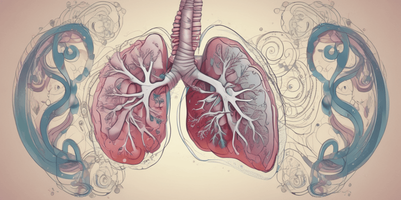
Pulmonary Disorder
- Obstructive diseases: pathological process that increases resistance to airflow, specifically expiratory airflow, due to luminal narrowing, loss of supporting structures surrounding airways, or constriction.
Asthma
- Chronic inflammatory condition with episodic obstruction of small and medium-sized bronchi
- Obstruction is reversible and inducible (Airway Hyperresponsive)
- Mechanism of Asthma:
- Exposure to trigger > Dendritic Cells take dust to other WBC like T-helper cell
- T cell sees this as dangerous and releases Cytokines
- B cells become activated causing production of IgE antibodies
- These antibodies attach to Mast cells, which develop surface proteins that retain memory of this specific allergen
- Rapid activation of Mast cells causes degradation, releasing leukotrienes and histamine
- Leukotrienes and histamine cause:
- Airway edema: Vasodilation and leakage of plasma into bronchial wall
- Mucus hypersecretion and plugging: Dilation of mucus glands
- Bronchial smooth muscle contraction: Parasympathetic activation
- Clinical significance of pathologic changes in Asthma:
- Airway narrowing decreases ventilation to some parts of the lungs > Decreased ventilation with normal perfusion (V/Q mismatch) > O2 will be decreased and CO2 will be increased
- Increased work of breathing
- Hypoxemia in severe asthma attack due to V/Q mismatch
- Presentation of asthma:
- Dyspnea (due to increased WOB)
- Wheezing (Increased Turbulence in narrow bronchi)
- Cough (Mucous hypersecretion)
- Hypoxemia in severe attack
- Tachypnea and tachycardia
- Hypocapnia
- Hypercapnia in severe and prolonged attacks
- Diagnosis:
- Pulmonary function test (spirometry)
- Decreased FVC, FEV1, and FEV1/FVC ratio (less than 0.7)
Chronic Obstructive Pulmonary Disease (COPD)
- Chronic progressive and irreversible obstructive disease caused by significant exposure to tobacco smoking or other environmental gases
- Mechanism of COPD:
- Medium-sized airway obstruction: Chronic Bronchitis
- Terminal bronchioles and alveolar destruction: Emphysema
- Clinical significance of pathologic changes in COPD:
- Airway obstruction and hyperinflations > leading to Increased WOB
- Impaired gas exchange > Hypoxemia, (+/-) Hypercapnia
- V/Q mismatch
- Pulmonary hypertension and right ventricular failure (cor pulmonale)
- Presentation of COPD:
- Dyspnea and exercise intolerance
- Productive cough (from inflamed airway)
- Wheezing
- Barrel-shaped chest (Increased anterior-posterior diameter)
- Pulmonary cachexia
- Accessory muscle use
- Pursed lip breathing
- Hypoxemia and +/- Hypercapnia
- RV failure signs (Peripheral edema, hepatosplenomegaly)
- Diagnosis of COPD:
- Pulmonary function test
- Decreased FVC, FEV1, and FEV1/FVC ratio (less than 0.7)
- NO or minimal increase in FEV1 post-bronchodilator
- Plethysmography: Increased FRC and Residual volume
- Chest X-ray: Hyperinflation and flattening of the diaphragm
Chronic Bronchitis
- Pathologic changes in airways:
- Exposure to irritants > Bronchial wall is infiltrated by monocytes and lymphocytes that secrete pro-inflammatory and proliferate substances
- Causing hypertrophy and hyperplasia of bronchial mucinous glands > Body responses
- Repairing of inflammation leading to fibrosis of bronchial wall..Bronchial remodeling occurs (Thickening of the wall and permanent narrowing of the lumen)
Emphysema
- Pathological changes in parenchyma:
- Exposure to irritant inducing inflammation > Immune cells will infiltrate
- Releasing lytic enzymes (Elastase, Collagenase)
- They will destroy Elastin and terminal bronchioles, alveoli creating large air pockets
- Loss of elastic fibers in the bronchial wall and surrounding tissues leads to airway obstruction, hyperinflation, and air trapping
Restrictive Diseases
- Pathological process that restricts lung expansion (compliance) particularly during inspiration
- Diseases:
- Intrapulmonary diseases (Interstitial lung disease, pulmonary edema)
- Extrapulmonary diseases (Pleural effusion, neuromuscular weakness, obesity, chest deformities)
Interstitial Lung Disease (ILD)
- Mechanism of ILD:
- Chronic diffuse inflammation of the alveolar interstitium > Thickening of interstitium from the accumulation of inflammatory cells > leading to repair inflammation causes deposition of fibers > diffused fibrosis of the interstitium
- Clinical significance of pathologic changes in ILD:
- Decreased lung compliance > Increased work of breathing
- Thickening alveolar capillary membrane > Diffusion problem > Slow diffusion of oxygen across the membrane > Hypoxia
- Hypoxemia > vasoconstriction > Pulmonary hypertension, right-sided heart failure (Cor Pulmonale)
- Presentation of ILD:
- Progressive dyspnea (Activity and then rest)
- Dry cough
- Progressive hypoxemia
- Digital clubbing
- S/S of RVF
- Diagnosis of ILD:
- Pulmonary function test
- Low FVC, low FEV1, but normal FEV1/FVC (0.7 or higher)
- Low RV and TLC
- X-ray: Diffuse reticular and nodular marking
Pleural Effusion
- Accumulation of fluids in the pleural space
- Causes:
- Increased production of pleural fluids
- Decreased drainage
- Mechanism of action:
- Transudation due to increased hydrostatic pressure: Hydrostatic pressure plasma out of the capillaries > Blood will fill pulmonary vessels > Back-up of pulmonary vessels and pleural vessels causing increased pressure > Forces fluid in pleural space > LOW PROTEIN
- Decreased oncotic pressure: Caused by liver cirrhosis or nephrotic syndrome > Osmotic pressure pulls fluid in that are protein-filled (albumin) > When there is a low amount of protein, oncotic pressure is low leading to fluids flowing out of capillaries > LOW PROTEIN
- Increased permeability of pleural capillaries seen in lung cancer, lung infection, and inflammatory conditions (lupus, RA)
- Presentation of pleural effusion:
- Asymptomatic (if less than 300ml)
- Pleuritic chest pain
- Dyspnea
- Decreased lung sounds
- Hypoxemia (compression of alveoli, decreased V/Q areas)
- Diagnosis of pleural effusion:
- Chemical analysis
- Transudate: Clear (serious). No protein and normal LDH
- Exudate: Cloudy (immune cells, bacteria, cancer cells), Increased protein and LDH
- X-ray: Blunting of costophrenic angle
Vascular Lung Disease
- Pathological process that changes resistance of pulmonary blood vessels
- NO changes in volume or airflow
Pulmonary Embolism (PE)
- Nearly all PE arise from deep veins of the legs above the knee (ileac, femoral, popliteal)
- Risk factors:
- Endothelial injury
- Stasis of blood: Immobility (bed rest)
- Hypercoagulopathy (thrombophilia)
- Pathological changes in PE:
- Pulmonary infarction
- Hypoxemia and hypocapnia
- Acute right ventricular and left ventricular failure (usually cause of death)
- Hypoxemia in PE:
- Embolus in the pulmonary artery > Leading to NO perfusion to ventilated alveoli (wasted/dead space) > No gas exchange occurs to this alveoli > Increased resistance to blood flow > Bloods diverted to areas with low resistance > Causing some areas to receive more blood (Q) than air (V) > Leading to VQ mismatch (Low V/Q areas > Hypoxemia with variable CO2 (normal or hypocapnic)
- Acute heart failure in PE:
- Occlusion of the pulmonary artery increases pulmonary vascular resistance > Increasing right ventricular afterload which makes right ventricles dilate and fail (acute cor pulmonale) > When the right ventricle dilates, the septum will push towards the left ventricles > Decreasing left ventricular filling leading to decreased left ventricular cardiac output causing acute left ventricular failure
- Presentation of PE:
- Acute onset of dyspnea
- Tachypnea, tachycardia
- Acute onset of pleuritic chest pain
- Hypoxemia
- Normal or reduced CO2
- Hypotension, syncope
- DVT
- Diagnosis of PE:
- CT angiography: Decreased pulmonary flow
Studying That Suits You
Use AI to generate personalized quizzes and flashcards to suit your learning preferences.
Description
This quiz covers pulmonary obstructive diseases, including asthma, COPD, and cystic fibrosis, and their effects on airflow and bronchial health. Learn about the pathological processes and symptoms of these conditions.


