Podcast
Questions and Answers
What is the primary function of the superior vena cava?
What is the primary function of the superior vena cava?
Which structure prevents backflow of blood into the right atrium during ventricular contraction?
Which structure prevents backflow of blood into the right atrium during ventricular contraction?
What is the primary role of the semilunar pulmonary valve?
What is the primary role of the semilunar pulmonary valve?
Which of the following structures has the thickest wall in the heart?
Which of the following structures has the thickest wall in the heart?
Signup and view all the answers
What is the role of the chordae tendineae in the heart?
What is the role of the chordae tendineae in the heart?
Signup and view all the answers
Where does the left atrium receive blood from?
Where does the left atrium receive blood from?
Signup and view all the answers
What characterizes the pectinate muscles in the right atrium?
What characterizes the pectinate muscles in the right atrium?
Signup and view all the answers
Which part of the heart helps to distribute oxygenated blood to the body?
Which part of the heart helps to distribute oxygenated blood to the body?
Signup and view all the answers
Which vein drains blood from the wall of the heart?
Which vein drains blood from the wall of the heart?
Signup and view all the answers
What is true about the right ventricle in comparison to the left ventricle?
What is true about the right ventricle in comparison to the left ventricle?
Signup and view all the answers
Study Notes
Cardiovascular System Anatomy
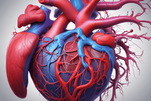
- The heart is a four-chambered muscular pump located in the middle mediastinum.
- It is positioned obliquely, with two-thirds on the left side and one-third on the right.
- The average heart size is comparable to a clenched fist.
- The average heart weight is 280 grams.
- The heart has upper atrial/venous chambers and lower ventricular/arterial chambers.
- Atria and ventricles are separated by coronary sulcus externally.
Heart Surfaces
- Sternocostal/Anterior Surface: Directed forwards and upwards, formed by the right atrium & auricle, part of the left auricle, anterior surface of the right ventricle (2/3), and anterior surface of the left ventricle (1/3).
- Diaphragmatic/Inferior Surface: Flat, facing upwards and backwards, formed by the two ventricles (2/3 left, 1/3 right). Lies over the central tendon of the diaphragm.
Heart Base
- Also known as the posterior surface.
- It is a quadrilateral shape.
- Formed by both atria (2/3rd left atrium, 1/3rd right atrium).
Heart Apex
- Conical area formed by the left ventricle.
- Directed downwards, forwards, and to the left.
- Located approximately 9 cm from the midline.
- In newborns, the apex beat is felt at the left 4th intercostal space.
Heart Borders
- Right Border: Formed by the right atrium, rounded and convex. Extends from the superior vena cava (SVC) to the inferior vena cava (IVC).
- Inferior Border: Sharp, extends from the IVC to the apex. Contains a notch called the incisura apicis cordis, closer to the apex.
- Left Border: Ill-defined, extends from the left auricle to the apex, separating the sternocostal surface from the left surface.
Heart Demarcations
- Heart is externally demarcated by grooves.
- Atrioventricular Groove: Surrounds the heart, except in front of the pulmonary trunk. Composed of anterior and posterior parts. Contains the right coronary artery, left circumflex branch of the left coronary artery, termination of great cardiac vein, and beginning of coronary sinus.
- Posterior Part of atrioventricular groove: Located between the base and diaphragmatic surface, Contains the coronary sinus and anastomosis of the right and left coronary arteries.
- Anterior Interventricular Groove: Runs downwards, forwards, and parallel to the left border, meeting the incisura apicis cordis. Indicates the anterior attachment of the interventricular septum, lodges the anterior interventricular artery, and great cardiac vein.
- Posterior Interventricular Groove: Runs forwards along the posterior surface, meeting the incisura apicis cordis. Indicates posterior attachment of interventricular septum. Lodges the posterior interventricular branch of the right coronary artery and middle cardiac vein.
Heart Wall
- Composed of epicardium (visceral pericardium), myocardium, and endocardium.
Right Atrium
- Has a muscular extension called the auricle.
- Interior of the anterior wall exhibits pectinate muscles.
- Has openings for the superior vena cava, inferior vena cava, and coronary sinus.
Right Ventricle
- Has a much thicker wall than the atria.
- Muscle projects into the cavity as papillary muscles, ending as chordae tendineae.
- Receives deoxygenated blood from the right atrium via the atrioventricular orifice.
Left Atrium
- Has a muscular extension called the auricle.
- Receives oxygenated blood from the lungs via four pulmonary veins.
- Pumps blood into the left ventricle through the left atrioventricular or bicuspid/mitral valve.
Left Ventricle
- Forms the apex of the heart.
- Its wall is three times thicker than the right ventricle.
- Muscle projects into the cavity as papillary muscles.
- Receives oxygenated blood from the left atrium via the bicuspid valve and ejects it into the aorta via the semilunar aortic valve.
Blood Supply to the Heart
- Supplied by the left and right coronary arteries, which branch from the ascending aorta.
- Left Coronary Artery (LCA): Branches to the left anterior descending (LAD) or anterior interventricular artery, the left marginal artery (LMA), and the left circumflex artery (Cx).
- Right Coronary Artery (RCA): Branches to the right marginal artery (RMA) and the posterior interventricular artery (Plv).
Coronary Veins
- Collect waste products from the myocardium.
- Drain into the coronary sinus.
- Major veins include the great cardiac vein, middle cardiac vein, small cardiac vein, and anterior cardiac vein.
Studying That Suits You
Use AI to generate personalized quizzes and flashcards to suit your learning preferences.
Related Documents
Description
Explore the intricate anatomy of the cardiovascular system, focusing on the heart's structure and surfaces. This quiz covers details such as the heart's chambers, positioning, and relation to other anatomical features, providing a comprehensive understanding for students of anatomy.


