Podcast
Questions and Answers
What is the role of the tricuspid valve in the heart?
What is the role of the tricuspid valve in the heart?
The tricuspid valve prevents backflow of blood from the right ventricle into the right atrium during ventricular contraction.
Describe the path of blood flow from the pulmonary arteries to the left atrium.
Describe the path of blood flow from the pulmonary arteries to the left atrium.
Blood flows from the pulmonary arteries to the lungs for oxygenation, then returns via the pulmonary veins to the left atrium.
What is the significance of papillary muscles and chordae tendineae in heart function?
What is the significance of papillary muscles and chordae tendineae in heart function?
Papillary muscles and chordae tendineae prevent the atrioventricular valves from inverting during ventricular contraction.
Identify the main vessel carrying oxygenated blood away from the heart and describe its location.
Identify the main vessel carrying oxygenated blood away from the heart and describe its location.
Signup and view all the answers
What differentiates the left ventricle from the right ventricle in terms of structure and function?
What differentiates the left ventricle from the right ventricle in terms of structure and function?
Signup and view all the answers
What is the primary role of the papillary muscles in the heart?
What is the primary role of the papillary muscles in the heart?
Signup and view all the answers
Describe the function of trabeculae carneae in the cardiac muscle.
Describe the function of trabeculae carneae in the cardiac muscle.
Signup and view all the answers
Explain the path of blood flow through the coronary arteries.
Explain the path of blood flow through the coronary arteries.
Signup and view all the answers
What anatomical structures delineate the septum between the right and left atria?
What anatomical structures delineate the septum between the right and left atria?
Signup and view all the answers
How do the aortic semilunar valves function in relation to systemic circulation?
How do the aortic semilunar valves function in relation to systemic circulation?
Signup and view all the answers
Study Notes
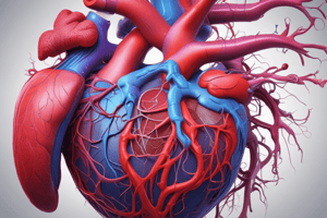
Heart Anatomy
- External Anterior View of the heart includes left atrium, left ventricle, right atrium, right ventricle.
- Left Atrium: Receives oxygenated blood from pulmonary veins; typically has two openings on each side.
- Right Ventricle: Features tricuspid valve (atrioventricular valve) and is responsible for pumping deoxygenated blood to pulmonary arteries.
-
Key Structures:
- Interventricular sulcus (anterior interventricular artery)
- Atrioventricular sulcus (contains R and L coronary arteries)
- Chordae tendineae attached to papillary muscles help control valve function.
- Trabeculae carneae are muscular ridges aiding in contraction and efficiency.
-
Major Blood Vessels:
- Superior vena cava and inferior vena cava bring deoxygenated blood to the right atrium.
- Pulmonary trunk branches into pulmonary arteries.
- Aorta carries oxygenated blood to the body.
Frontal Cross-Section
- Right Atrium: Contains openings for superior vena cava, inferior vena cava, and coronary sinus; important for blood return to the heart.
- Interatrial Septum: Separates left and right atria, includes fossa ovalis.
- Pectinate Muscles: Present in the right atrium, provide structural support.
Coronary Circulation
- Purpose: Supplies the heart muscle with oxygenated blood; shortest circulation in the body.
-
Arterial Supply:
- Left and right coronary arteries arise from the aorta's base.
- Left coronary artery includes the anterior interventricular artery and circumflex artery, which anastomoses with the right coronary artery's posterior interventricular artery.
- Right coronary artery gives rise to marginal artery and continues to the posterior side for blood supply.
- Venous Return: Blood collected by cardiac veins (small, middle, great) drains into the coronary sinus, opening into the right atrium.
Clinical Considerations
- Blockage in Coronary Arteries: Can lead to myocardium damage, causing heart tissue death (ischemia).
- Need for Cardiac Circulation: Heart requires its own blood supply to sustain muscle function and prevent ischemic conditions.
Blood Flow Through the Heart
- Discussion of blood flow mechanics is essential for understanding overall cardiovascular function.
- Use heart models to visualize and comprehend the journey of blood through chambers and vessels.
Studying That Suits You
Use AI to generate personalized quizzes and flashcards to suit your learning preferences.
Description
Explore the critical roles of various heart components such as the tricuspid valve, pulmonary arteries, and the importance of papillary muscles and chordae tendineae. Understand the distinction between the left and right ventricles in terms of structure and function, and learn about the main vessel carrying oxygenated blood from the heart. This quiz will deepen your knowledge of cardiovascular anatomy.