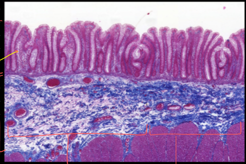
What structures are visible in the attached histological image?

Understand the Problem
The question is likely asking for an analysis or identification of the structures visible in the histological image, which seems to depict a cross-section of a tissue sample.
Answer
Villi, crypts, mucosa, submucosa, muscularis.
The image shows villi, crypts, and layers of tissue such as the mucosa, submucosa, and muscularis.
Answer for screen readers
The image shows villi, crypts, and layers of tissue such as the mucosa, submucosa, and muscularis.
More Information
The histological image appears to be a section through intestinal tissue, revealing key structures like the finger-like villi, which increase surface area for absorption, and the underlying layers of tissue.
Tips
Be careful to correctly identify tissue layers and structures by their staining patterns and morphology.
AI-generated content may contain errors. Please verify critical information