Podcast
Questions and Answers
Which magnification level would provide detailed observations of eye structures?
Which magnification level would provide detailed observations of eye structures?
What is the purpose of using a broad beam in slit-lamp examination?
What is the purpose of using a broad beam in slit-lamp examination?
What is typically assessed when examining the cornea and iris?
What is typically assessed when examining the cornea and iris?
At what angle is tangential illumination best utilized during eye examination?
At what angle is tangential illumination best utilized during eye examination?
Signup and view all the answers
Which condition is indicated by aqueous flares in the anterior chamber?
Which condition is indicated by aqueous flares in the anterior chamber?
Signup and view all the answers
Which part of the slit-lamp is primarily used to adjust the focus and power of the magnification?
Which part of the slit-lamp is primarily used to adjust the focus and power of the magnification?
Signup and view all the answers
What is the correct order of anterior segment examination starting from the lids and lashes?
What is the correct order of anterior segment examination starting from the lids and lashes?
Signup and view all the answers
Which illumination technique is best suited for evaluating corneal thickness and depth of features?
Which illumination technique is best suited for evaluating corneal thickness and depth of features?
Signup and view all the answers
What type of filter is used in diffuser lighting to reduce glare without affecting color perception?
What type of filter is used in diffuser lighting to reduce glare without affecting color perception?
Signup and view all the answers
Which slit-lamp illumination technique is specifically indicated for detecting aqueous flare and cell debris?
Which slit-lamp illumination technique is specifically indicated for detecting aqueous flare and cell debris?
Signup and view all the answers
Study Notes
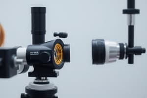
Slit-Lamp Biomicroscope Components
- Mechanical Support: Forehead rest, chin rest, fixation target, power supply unit, locking controls.
- Observation System: Binocular eyepieces, camera/video adaptor, observation tube (on some models), magnification changer.
- Illumination System: Lamp housing unit, slit width and height control, neutral density filter, cobalt blue light, red-free (green) filter, field size control, diffuser, prism.
Anterior Segment Examination Sequence
- Lids and lashes
- Conjunctiva
- Cornea
- Tear film
- Eyelid eversion
- Anterior chamber
- Angle of the anterior chamber
- Iris
- Crystalline lens
- Anterior vitreous
Slit-Lamp Illumination Techniques & Applications
- Diffused Illumination: Low to high magnification, neutral density filter, moderate slit height; used to examine lids, lashes, conjunctiva, sclera, iris, pupil. Assesses blepharitis, trichiasis, conjunctival concretions, pterygium, pinguecula.
- Parallel Epiped Illumination: ~30° angle, medium to high magnification, neutral density filter, variable slit width (2-3 mm); used to assess corneal stroma (vacuoles, microcysts, dystrophies, striae, folds), lens surface, endothelium, nerve fibers, blood vessels, infiltrates. Evaluates corneal thickness.
- Direct Focal Illumination: Medium to high magnification, neutral density filter, narrow beam; used for evaluating corneal thickness, identifying variations in corneal curvature, angle closure glaucoma, nerve fibers, blood vessels, infiltrates, cataracts.
- Optic Section Illumination: 60° angle, medium to high magnification, neutral density filter, full slit height; used to assess depth of visible features within the cornea.
- Conical Beam Illumination: >50°, medium to high magnification, neutral density filter, small circular slit; used to examine anterior chamber (aqueous flare, pigmentation, cell debris), corneal layers (vascularization, epithelial edema, microcysts), crystalline lens (vacuoles, dystrophies, opacities, contact lens deposits).
- Retro-illumination: Coaxial (~0.5°), medium to high magnification, neutral density filter, small circular slit; assesses localized epithelial edema (central corneal clouding), lens opacities, aqueous flares.
- Indirect Sclerotic Scatter Illumination: Low to medium magnification, neutral density filter, broad beam, slit width (2-3 mm); used to assess corneal scars, foreign bodies in the cornea, and endothelial blebs.
- Specular Reflection: High magnification, neutral density filter, broad beam, slit width (2-3 mm), 25°/30° angle; used to evaluate endothelial cell layer, tear film debris, and tear film lipid layer thickness.
- Tangential Illumination: Low to medium magnification, neutral density filter, wide open slit, 70°-80° angle; assesses general integrity of cornea and iris, iris freckles, and tumors.
- Oscillatory Beam Illumination: Low to medium magnification, neutral density filter, medium to broad beam, ~60°, variable height; evaluates lens opacities, aqueous flares.
Slit-Lamp Magnification and Observed Area
- Low Magnification (7X - 10X): General eye examination.
- Medium Magnification (20X - 25X): Examination of structure layers.
- High Magnification (30X - 40X): Detailed examination.
Studying That Suits You
Use AI to generate personalized quizzes and flashcards to suit your learning preferences.
Related Documents
Description
Test your knowledge on the components and examination techniques of the slit-lamp biomicroscope. This quiz covers essential parts of the equipment, the sequence of anterior segment examinations, and various illumination techniques. Perfect for students and professionals in ophthalmology.


