Podcast
Questions and Answers
What condition is characterized by abnormal additional eyelash lines?
What condition is characterized by abnormal additional eyelash lines?
- Entropian
- Trichiasis
- Distichiasis (correct)
- Madarosis
Which of the following is NOT a method of assessing the tear film?
Which of the following is NOT a method of assessing the tear film?
- Non-Invasive Tear Break-Up-Time (NIBUT)
- Tear prism height
- Schirmer test (correct)
- Fluorescein staining
Which condition involves the inward turning of the eyelashes?
Which condition involves the inward turning of the eyelashes?
- Entropian (correct)
- Ptosis
- Ectropian
- Trichiasis
What is the primary focus when examining the meibomian glands during a visual examination?
What is the primary focus when examining the meibomian glands during a visual examination?
In a slit-lamp examination, which anatomical structure is evaluated LAST?
In a slit-lamp examination, which anatomical structure is evaluated LAST?
Which of the following structures is evaluated for hyperemia during a slit-lamp examination?
Which of the following structures is evaluated for hyperemia during a slit-lamp examination?
What is observed when evaluating the bulbar conjunctiva?
What is observed when evaluating the bulbar conjunctiva?
During a slit-lamp examination, what is the difference between a parallelepiped and an optic section?
During a slit-lamp examination, what is the difference between a parallelepiped and an optic section?
Which part of the eye should be examined last during a thorough slit-lamp examination?
Which part of the eye should be examined last during a thorough slit-lamp examination?
The presence of which feature on the palpebral conjunctiva may indicate chronic allergic reaction?
The presence of which feature on the palpebral conjunctiva may indicate chronic allergic reaction?
What is the estimated angle range for a slit beam considered closed?
What is the estimated angle range for a slit beam considered closed?
During the OSCE, how many techniques must be performed within the 12 minutes procedure?
During the OSCE, how many techniques must be performed within the 12 minutes procedure?
Which ratio indicates a higher likelihood of angle closure?
Which ratio indicates a higher likelihood of angle closure?
What is one requirement during the OSCE regarding findings?
What is one requirement during the OSCE regarding findings?
What is the estimated angle range indicating that angle closure is likely?
What is the estimated angle range indicating that angle closure is likely?
Which technique is NOT used in indirect illumination during a slit-lamp examination?
Which technique is NOT used in indirect illumination during a slit-lamp examination?
What is the correct focusing requirement for an emmetrope during a slit-lamp examination?
What is the correct focusing requirement for an emmetrope during a slit-lamp examination?
When assessing the anterior chamber angle during a slit-lamp examination, which illumination technique is primarily employed?
When assessing the anterior chamber angle during a slit-lamp examination, which illumination technique is primarily employed?
What is critical to maintain image focus while using a slit lamp?
What is critical to maintain image focus while using a slit lamp?
What should be done after decoupling the slit lamp?
What should be done after decoupling the slit lamp?
Which of the following structures is examined first when conducting a slit-lamp examination from outside to inside?
Which of the following structures is examined first when conducting a slit-lamp examination from outside to inside?
How should the illumination arm be moved during an anterior eye assessment?
How should the illumination arm be moved during an anterior eye assessment?
What is the recommended action after a slit-lamp examination to maintain cleanliness in the examination area?
What is the recommended action after a slit-lamp examination to maintain cleanliness in the examination area?
What is the main purpose of using a parallelepiped in corneal assessment?
What is the main purpose of using a parallelepiped in corneal assessment?
What causes microcysts formation in the corneal epithelium?
What causes microcysts formation in the corneal epithelium?
How is corneal edema characterized at 12% swelling?
How is corneal edema characterized at 12% swelling?
Which term describes vertically running densifications in the stromal tissue?
Which term describes vertically running densifications in the stromal tissue?
What does Van Herick's grading primarily evaluate?
What does Van Herick's grading primarily evaluate?
What causes guttata formation in the endothelium?
What causes guttata formation in the endothelium?
Which of the following findings would indicate the presence of excessive corneal swelling?
Which of the following findings would indicate the presence of excessive corneal swelling?
Which examination method helps detect changes in the endothelium layer?
Which examination method helps detect changes in the endothelium layer?
What distinguishes Descemet's folds from striae?
What distinguishes Descemet's folds from striae?
Which finding correlates with chronic hypoxia in the endothelium?
Which finding correlates with chronic hypoxia in the endothelium?
Flashcards
Ptosis
Ptosis
drooping of the upper eyelid
Entropian
Entropian
Turning inward of the eyelid margin
Ectropian
Ectropian
Turning outward of the eyelid margin
Distichiasis
Distichiasis
Signup and view all the flashcards
Trichiasis
Trichiasis
Signup and view all the flashcards
Direct illumination
Direct illumination
Signup and view all the flashcards
Sclerotic scatter
Sclerotic scatter
Signup and view all the flashcards
Retro illumination
Retro illumination
Signup and view all the flashcards
Conjunctiva
Conjunctiva
Signup and view all the flashcards
Cornea
Cornea
Signup and view all the flashcards
Anterior chamber
Anterior chamber
Signup and view all the flashcards
Iris
Iris
Signup and view all the flashcards
Crystalline lens
Crystalline lens
Signup and view all the flashcards
Pinguecula
Pinguecula
Signup and view all the flashcards
Pterygium
Pterygium
Signup and view all the flashcards
Conjunctival Hyperemia
Conjunctival Hyperemia
Signup and view all the flashcards
Limbal Injection
Limbal Injection
Signup and view all the flashcards
Optic Section
Optic Section
Signup and view all the flashcards
Parallelepiped Examination
Parallelepiped Examination
Signup and view all the flashcards
Optic Section Examination
Optic Section Examination
Signup and view all the flashcards
Specular Reflection Examination
Specular Reflection Examination
Signup and view all the flashcards
Microcysts
Microcysts
Signup and view all the flashcards
Vacuoles
Vacuoles
Signup and view all the flashcards
Corneal Stroma Oedema
Corneal Stroma Oedema
Signup and view all the flashcards
Striae
Striae
Signup and view all the flashcards
Descemet's Folds
Descemet's Folds
Signup and view all the flashcards
Polymegathism
Polymegathism
Signup and view all the flashcards
VanHerick's Grading
VanHerick's Grading
Signup and view all the flashcards
What is the angle of the anterior chamber?
What is the angle of the anterior chamber?
Signup and view all the flashcards
What is angle closure?
What is angle closure?
Signup and view all the flashcards
What is a narrow angle?
What is a narrow angle?
Signup and view all the flashcards
What is gonioscopy?
What is gonioscopy?
Signup and view all the flashcards
What is a gonioscope?
What is a gonioscope?
Signup and view all the flashcards
Study Notes
Lecture Recording Information
- The lecture is being recorded as part of Plymouth University's Content Capture Project
- The recording will be available via the Panopto block on the module DLE pages
- Students can ask questions during the lecture
- Comments may appear on the recording
- Option to pause the recording if a question is posed that the lecturer doesn't want on the recording
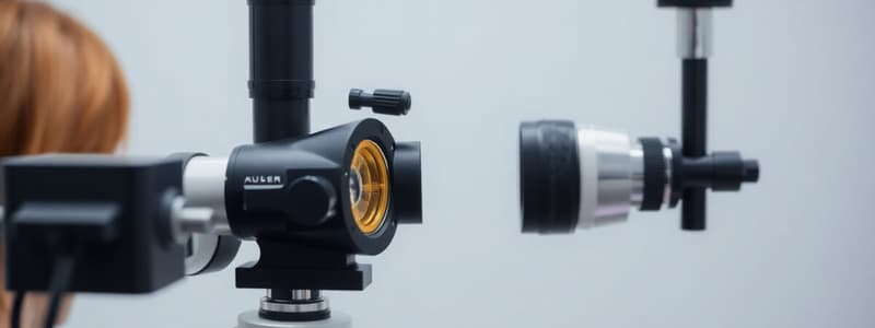
Optometry Skills - Slit Lamp Biomicroscopy 2
- Course Code: OPT506
- Lecturer: Dr. Asma Zahidi
- One-time code: HC-YK-UK
Practical Session Feedback
- Wipe down the slit lamp
- Focusing: ±0.50D emmetropic/fully corrected
- Use joystick to focus on different structures
- Maintain image focus by keeping hand on joystick
- Use both hands to operate the slit lamp
- Couple the slit lamp after decoupling
- Leave the bay tidy
Slit Lamp Examination Flowchart
- White light: Lid, margins/eyelashes, tears/tear film, Conjunctiva (Bulbar, Palpebral), Limbus, Cornea, Anterior chamber, Iris, Crystalline lens
- Blue light + NaFl: Tears/tear film, Cornea, Conjunctiva (Bulbar, Palpebral)
Assessment of the Tear Film
- Non-invasive methods:
- Tear prism height
- Non-invasive Tear Break-up Time (NIBUT)
- Lipid layer evaluation (pages 109-110)
Assessment of the Conjunctiva
- White light:
- Bulbar (lumps/bumps, pinguecula, pterygium, hyperemia)
- Limbus (hyperemia, limbal injection)
- Palpebral (*leave upper palpebral conjunctiva after instilled NaFl - lumps/bumps (papillae, concretions))
Parallelepiped vs. Optic Section
- Parallelepiped: Location on the surface of the eye
- Optic section: Depth/layer of the eye
Assessment of the Cornea
- Parallelepiped (medium mag):
- Examines cornea texture and precorneal tear film
- Identifies scars, irregularities, and tear film mobility post-blink
- Sclerotic scatter: Identifies scars, deposits, edema, and abnormal scattered light
- Optic section (medium/high mag): Assesses the curvature, thickness, and depth of changes/scars on the cornea
- Specular reflection: Detects changes in the endothelium layer
Epithelium - Microcysts and Vacuoles
- Microcysts: Caused by oxygen deficiency inducing metabolism suppression
- Vacuoles: Caused by epithelial edema, superficial keratitis, trauma, or inherited epithelial dystrophy
- Differential: Microcysts have opposite refraction, vacuoles have concurrent refraction
Stroma - Oedema
- Cause: Fluid accumulation between cells
- Findings: Subjective (streaking, foggy vision, fluctuations) and objective (classification using striae and Descemet folds)
- Levels: 0% (no striae), 5% (single striae), 7% (several striae), 12% (striae & folds), 16% (striae, folds, microcysts, vacuoles)
Stroma - Striae
- Vertically running densifications in the stromal tissue without ramification (delimitation to nerve fibre)
Stroma - Descemet's folds
- Horizontal, evenly spaced, fine, whitish lines with double contours
Endothelium
- Key features: Polymorphism, Polymegathism, Bleps, Guttata
Anterior Chamber Assessment
- Assessment method using Van Herick's grading (Grade 4-0)
Iris Assessment
Crystalline Lens Assessment
OSCE 1 - Summative
- Assigned to a bay: Students are assigned a specific area
- Assessor: An assessor is at the student's bay
- Fellow student: Serves as a patient
- 1 minute reading time: For instructions
- Instruction sheet: Contains 5 techniques to perform
- One eye: Students perform the techniques on one eye only
- Eye selection: The assessor will inform the student which eye to use
OSCE Station 1
- Duration: 12 minutes
- Techniques: 3 basic, 2 advanced
- Communication: Explain each step
- Observation: Clearly show the technique and targeted structures
- Recording: No recording of findings is necessary
Studying That Suits You
Use AI to generate personalized quizzes and flashcards to suit your learning preferences.


