Podcast
Questions and Answers
ACh is produced in the nerve terminal and stored in ______.
ACh is produced in the nerve terminal and stored in ______.
synaptic vesicles
After release, ACh binds to receptors on ______.
After release, ACh binds to receptors on ______.
Muscle
Type I muscle fibers are also known as ______ twitch fibers.
Type I muscle fibers are also known as ______ twitch fibers.
slow
Type II muscle fibers are also known as ______ twitch fibers.
Type II muscle fibers are also known as ______ twitch fibers.
Signup and view all the answers
The NMJ is also known as a __________ synapse.
The NMJ is also known as a __________ synapse.
Signup and view all the answers
The contractile unit of skeletal muscle is called a ______.
The contractile unit of skeletal muscle is called a ______.
Signup and view all the answers
The neurotransmitter released at the NMJ is __________.
The neurotransmitter released at the NMJ is __________.
Signup and view all the answers
Actin and Myosin give muscle a striated ______.
Actin and Myosin give muscle a striated ______.
Signup and view all the answers
The sarcoplasmic reticulum serves as a ______ storehouse.
The sarcoplasmic reticulum serves as a ______ storehouse.
Signup and view all the answers
The __________ motor neuron is also referred to as the alpha motor neuron.
The __________ motor neuron is also referred to as the alpha motor neuron.
Signup and view all the answers
The ______ cross-bridge cycle explains the mechanisms of muscle contraction.
The ______ cross-bridge cycle explains the mechanisms of muscle contraction.
Signup and view all the answers
There is a synaptic cleft of about __________ micrometers at the NMJ.
There is a synaptic cleft of about __________ micrometers at the NMJ.
Signup and view all the answers
The muscle action potential is initiated when ACh binds to __________ on the muscle.
The muscle action potential is initiated when ACh binds to __________ on the muscle.
Signup and view all the answers
Skeletal muscle fibers are organized into ______ that consist of actin and myosin.
Skeletal muscle fibers are organized into ______ that consist of actin and myosin.
Signup and view all the answers
Mitochondria in muscle fibers are responsible for producing ______.
Mitochondria in muscle fibers are responsible for producing ______.
Signup and view all the answers
The sequence of events that occurs when a skeletal muscle contracts is known as __________ formation.
The sequence of events that occurs when a skeletal muscle contracts is known as __________ formation.
Signup and view all the answers
The area of the neuron that releases neurotransmitters is called the __________ terminal.
The area of the neuron that releases neurotransmitters is called the __________ terminal.
Signup and view all the answers
__________ are numerous in both the presynaptic and postsynaptic terminals at the NMJ.
__________ are numerous in both the presynaptic and postsynaptic terminals at the NMJ.
Signup and view all the answers
The region between the motor nerve and the muscle fiber is known as the __________ cleft.
The region between the motor nerve and the muscle fiber is known as the __________ cleft.
Signup and view all the answers
An __________ motor neuron is responsible for transmitting signals to skeletal muscle.
An __________ motor neuron is responsible for transmitting signals to skeletal muscle.
Signup and view all the answers
Study Notes
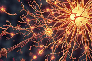
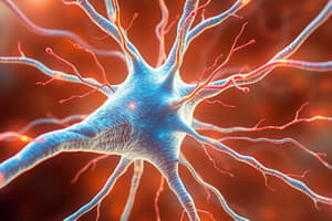
Neuromuscular Junction (NMJ)
- The NMJ is the synapse between a motor neuron and a muscle fiber.
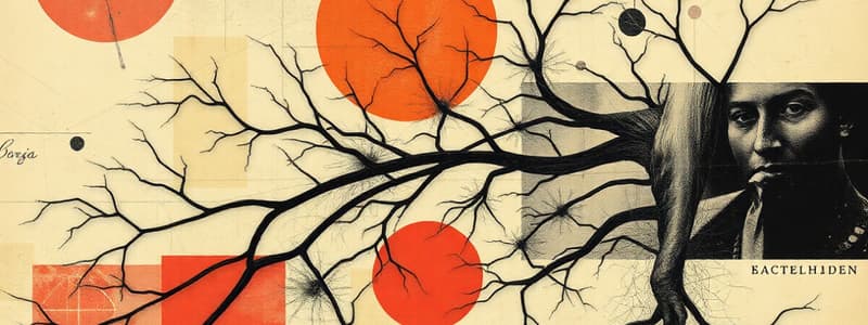
- Alpha motor neurons are also known as lower motor neurons and are responsible for stimulating muscle contraction.
- The synaptic cleft is the space between the motor neuron and the muscle fiber.
- Acetylcholine (ACh) is the neurotransmitter released at the NMJ.
Structure of the NMJ
- The motor neuron terminal has a presynaptic terminal and the muscle fiber has a postsynaptic terminal.
- The synaptic cleft is about 20 µm wide, ensuring no physical contact between the nerve and the muscle.
- The presynaptic terminal has many mitochondria for ATP production needed for neurotransmitter release.
- The postsynaptic terminal contains receptors for the neurotransmitter.
Neurotransmitter Release at the NMJ
- ACh is stored in synaptic vesicles in the presynaptic terminal.
- The arrival of an action potential at the presynaptic terminal triggers the release of ACh via exocytosis.
- ACh diffuses across the synaptic cleft and binds to receptors on the muscle fiber membrane.
Events after Neurotransmitter Release
- Binding of ACh to its receptors on the muscle fiber membrane opens ion channels, leading to depolarization of the muscle fiber membrane.
- This depolarization generates an end-plate potential (EPP) which triggers an action potential in the muscle fiber.
- The muscle fiber action potential travels along the sarcolemma, leading to muscle contraction.
- ACh is rapidly broken down by acetylcholinesterase (AChE) in the synaptic cleft, ending the signal and preventing continuous muscle contraction.
Types of Skeletal Muscle Fibers
-
Type I (Slow Twitch) Muscle Fibers (Red):
- Used for postural muscles
- High myoglobin content
- Many mitochondria for oxidative metabolism
- Fatigue-resistant
-
Type II (Fast Twitch) Muscle Fibers (White):
- Found in extraocular muscles
- Lower myoglobin content
- Fewer mitochondria
- Use glycolytic metabolism for energy production
- More fatigue-prone
Gross Structure of Muscle
- Muscle fibers are composed of myofibrils, which are in turn made up of myofilaments of actin and myosin.
- The arrangement of actin and myosin filaments gives skeletal muscle a striated appearance.
Organization of Skeletal Muscle Fibers
- Myofibrils are made up of repeating contractile units called sarcomeres.
- The sarcomere is the basic unit of muscle contraction.
- T-tubules allow the propagation of the muscle action potential throughout the muscle fiber.
- Sarcoplasmic reticulum (SR) is a specialized endoplasmic reticulum in muscle fibers that stores and releases calcium ions for muscle contraction.
- The SR is connected to the T-tubules.
Histology of Skeletal Muscle Fibers
- The thin filaments are made of the protein actin.
- The thick filaments are made of the protein myosin.
- Titin is a large protein that connects myosin to the Z-disc.
- Troponin and tropomyosin are proteins that regulate actin-myosin interaction during muscle contraction.
The Contraction-Relaxation Cycle (Cross-Bridge Formation)
-
Muscle contraction:
- Calcium ions released from the SR bind to troponin.
- This causes a conformational change in tropomyosin, exposing the myosin-binding site on actin.
- Myosin heads attach to the actin filament (cross-bridge formation).
- The myosin heads swivel, pulling the actin filament towards the center of the sarcomere.
- ATP is hydrolyzed, providing energy for the power stroke .
- The myosin heads detach from the actin filament, and the cycle repeats.
-
Muscle relaxation:
- Calcium ions are pumped back into the SR by active transport.
- Troponin returns to its original conformation.
- Tropomyosin covers the myosin-binding sites on actin.
- Myosin can no longer bind to actin, and the muscle relaxes.
Studying That Suits You
Use AI to generate personalized quizzes and flashcards to suit your learning preferences.
Related Documents
Description
Explore the structure and function of the neuromuscular junction (NMJ) in this quiz. Learn about the roles of alpha motor neurons, acetylcholine release, and the components of the NMJ. Test your knowledge on how these elements work together to stimulate muscle contractions.