Podcast
Questions and Answers
What is the primary function of acetylcholine (ACh) after it is released into the synaptic cleft?
What is the primary function of acetylcholine (ACh) after it is released into the synaptic cleft?
Which type of skeletal muscle fiber is characterized by a high myoglobin content and resistance to fatigue?
Which type of skeletal muscle fiber is characterized by a high myoglobin content and resistance to fatigue?
What is the structural significance of the sarcomere in skeletal muscle contraction?
What is the structural significance of the sarcomere in skeletal muscle contraction?
What role does the sarcoplasmic reticulum (SR) play in muscle contraction?
What role does the sarcoplasmic reticulum (SR) play in muscle contraction?
Signup and view all the answers
Which of the following best describes Type II muscle fibers?
Which of the following best describes Type II muscle fibers?
Signup and view all the answers
What is the primary composition of a sarcomere?
What is the primary composition of a sarcomere?
Signup and view all the answers
What is the function of acetylcholinesterase (AChE) following the action of acetylcholine?
What is the function of acetylcholinesterase (AChE) following the action of acetylcholine?
Signup and view all the answers
Which part of the muscle fiber is responsible for the propagation of action potentials?
Which part of the muscle fiber is responsible for the propagation of action potentials?
Signup and view all the answers
What distinguishes Type I muscle fibers from Type II fibers in terms of energy metabolism?
What distinguishes Type I muscle fibers from Type II fibers in terms of energy metabolism?
Signup and view all the answers
What characterizes the neuromuscular junction (NMJ) compared to other types of synapses?
What characterizes the neuromuscular junction (NMJ) compared to other types of synapses?
Signup and view all the answers
What is the primary role of acetylcholine (ACh) at the NMJ?
What is the primary role of acetylcholine (ACh) at the NMJ?
Signup and view all the answers
Which component is NOT part of a muscle fiber's structure?
Which component is NOT part of a muscle fiber's structure?
Signup and view all the answers
What happens after acetylcholine binds to its receptors on the muscle?
What happens after acetylcholine binds to its receptors on the muscle?
Signup and view all the answers
Which is NOT a characteristic of the alpha motor neuron?
Which is NOT a characteristic of the alpha motor neuron?
Signup and view all the answers
Which sequence correctly describes the process that occurs at the NMJ?
Which sequence correctly describes the process that occurs at the NMJ?
Signup and view all the answers
What is a notable feature of the presynaptic terminal at the NMJ?
What is a notable feature of the presynaptic terminal at the NMJ?
Signup and view all the answers
Which event occurs following neurotransmitter release into the synaptic cleft?
Which event occurs following neurotransmitter release into the synaptic cleft?
Signup and view all the answers
Why is the synaptic cleft important in neuromuscular transmission?
Why is the synaptic cleft important in neuromuscular transmission?
Signup and view all the answers
Study Notes
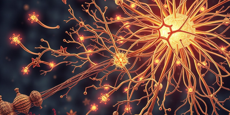
Neuromuscular Junction (NMJ)
- The NMJ is an example of a synapse where a motor nerve meets a muscle.
- The nerve and muscle don't physically touch, but are separated by a synaptic cleft of about 20um.
- The transfer of messages occurs via the release of neurotransmitter acetylcholine (ACh) into the synaptic cleft.
- ACh is produced in the nerve terminal and stored in synaptic vesicles.
- When released, ACh binds to receptors on the muscle, triggering an end-plate potential (EPP) in the muscle cell.
- This initiates an action potential in the skeletal muscle, causing it to contract.
- ACh is then broken down by acetylcholinesterase (AChE), and its components are recycled.
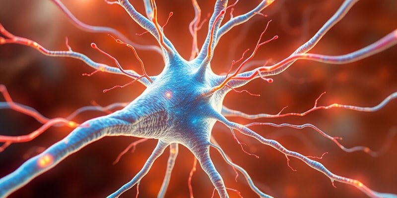
Alpha Motor Neuron
- Also known as a lower motor neuron.
- Responsible for transmitting signals from the brain to muscle fibers.
- The axon of the alpha motor neuron terminates at the NMJ.
Structure of the NMJ
- Consists of a presynaptic terminal, synaptic cleft, and a postsynaptic terminal.
- The presynaptic terminal is the end of the motor neuron axon, containing synaptic vesicles filled with ACh.
- The synaptic cleft is the space between the presynaptic and postsynaptic terminals.
- The postsynaptic terminal is the muscle fiber membrane, with receptors for ACh.
Muscle Fiber Types
-
Type I (Slow Twitch):
- Also known as red muscle fibers.
- Found in postural muscles, responsible for sustained, low-force contractions.
- High in myoglobin and mitochondria, indicating high oxidative metabolism.
- Fatigue-resistant.
-
Type II (Fast Twitch):
- Also known as white muscle fibers.
- Found in extraocular muscles, used for rapid, powerful contractions.
- Lower myoglobin and mitochondria, indicating lower oxidative metabolism and higher glycolytic metabolism.
- Fatigue-prone.
Muscle Fiber Structure
- Muscle fibers contain myofibrils, which are composed of myofilaments of actin and myosin, giving the muscle a striated appearance.
- Myofibrils are enveloped by a network of T-tubules that help spread the muscle action potential.
- Contain large quantities of mitochondria for ATP production and a sarcoplasmic reticulum for calcium storage.
Sarcomere
- The contractile unit of skeletal muscle.
- Extends from one Z disc to the next, creating the striated appearance.
- Composed of actin (thin filament), myosin (thick filament), and other proteins like titin, troponin, and tropomyosin.
Contraction-Relaxation Cycle (Cross-Bridge Formation)
- During contraction, the sarcomere shortens, and the Z-lines move closer together.
- This occurs due to the sliding of actin filaments over myosin filaments.
- Myosin heads bind to actin, forming cross-bridges.
- ATP hydrolysis provides energy for myosin head movement, pulling actin filaments towards the center of the sarcomere.
- During relaxation, calcium is pumped back into the sarcoplasmic reticulum, causing myosin heads to detach from actin, allowing the sarcomere to lengthen.
Studying That Suits You
Use AI to generate personalized quizzes and flashcards to suit your learning preferences.
Related Documents
Description
This quiz covers the neuromuscular junction (NMJ), a critical synapse where motor nerves communicate with muscles. Learn about the role of acetylcholine, the structure of the NMJ, and the function of alpha motor neurons in muscle contraction. Test your understanding of these fundamental concepts in neuromuscular physiology.