Podcast
Questions and Answers
What are the two main parts of the cerebrum?
What are the two main parts of the cerebrum?
Diencephalon and telencephalon
Which of the following structures is the largest commissure in the brain?
Which of the following structures is the largest commissure in the brain?
The occipital lobe is located anterior to the first imaginary line.
The occipital lobe is located anterior to the first imaginary line.
False (B)
Match the following lobes of the cerebrum with their corresponding locations.
Match the following lobes of the cerebrum with their corresponding locations.
Signup and view all the answers
The part of the cerebral cortex that guides motor functions is called the _______.
The part of the cerebral cortex that guides motor functions is called the _______.
Signup and view all the answers
What are gyri and sulci?
What are gyri and sulci?
Signup and view all the answers
What is the main function of the sensory area of the cerebral cortex?
What is the main function of the sensory area of the cerebral cortex?
Signup and view all the answers
The parahippocampal gyrus is part of the archicortex.
The parahippocampal gyrus is part of the archicortex.
Signup and view all the answers
What area corresponds to the auditory area of the cerebral cortex?
What area corresponds to the auditory area of the cerebral cortex?
Signup and view all the answers
What happens when there is damage to area 4 of Brodmann?
What happens when there is damage to area 4 of Brodmann?
Signup and view all the answers
The bundle of fibers that interconnects identical areas of both cerebral hemispheres is called the _______.
The bundle of fibers that interconnects identical areas of both cerebral hemispheres is called the _______.
Signup and view all the answers
Study Notes
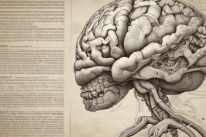
Overview of the Cerebrum
- Largest brain part located in the anterior and middle cranial fossae.
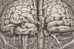
- Comprised of two major parts: diencephalon (central core) and telencephalon (cerebral hemispheres), separated by the longitudinal cerebral fissure.
- The fissure contains the falx cerebri and anterior cerebral arteries and hosts the corpus callosum, which connects hemispheres.
Poles and Borders of the Cerebrum
- Recognized poles: frontal pole (anterior), temporal pole (between frontal and occipital), occipital pole (posterior).
- Each cerebral hemisphere has three borders: superomedial, inferolateral, and inferomedial.
- The inferomedial border is further divided into the medial orbital border (anterior) and the medial occipital border (posterior).
Surfaces of the Cerebrum
- The hemispheres have three primary surfaces: superolateral, medial, and inferior surfaces.
- The inferior surface is subdivided into an anterior orbital part and a posterior tentorial part.
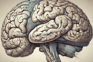
Lobes of the Cerebrum
- The lobes are distinguished by key sulci, including the posterior ramus of the lateral sulcus and the central sulcus for localization.
- Frontal lobe: Anterior to the central sulcus; includes areas for higher cognitive functions.
- Parietal lobe: Behind the central sulcus; handles sensory information and spatial awareness.
- Temporal lobe: Below the lateral sulcus; involved in processing auditory information and memory.
- Occipital lobe: Behind the parieto-occipital sulcus; primarily responsible for visual processing.
Gyri and Sulci
- Gyri are ridges, and sulci are grooves of the cerebral cortex; collectively known as Gyrencephalic brain in higher mammals.
- Frontal lobe features include the precentral sulcus and division into superior, middle, and inferior frontal gyri.
- Temporal lobe contains superior and inferior temporal sulci dividing it into respective gyri.
- Parietal lobe is segmented by the postcentral sulcus and intraparietal sulcus into superior and inferior parietal lobules.
Medial Surface Features
- Significant sulci include the cingulate sulcus, dividing the gyrus cinguli from the corpus callosum.
- The medial surfaces contain the paracentral lobule and the cuneus, defined by the parieto-occipital and calcarine sulci.
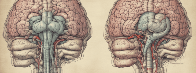
Inferior Surface Characteristics
- Inflected by the olfactory sulcus, associated with olfactory tracts, and further detailed into gyri and sulci like the lingual gyrus and uncus.
Cerebral Cortex Overview
- The highly evolved cerebral cortex consists of neurons and neuroglia, comprising about 14 billion neurons with layered organization.
- It includes three divisions based on evolutionary significance: archicortex (primitive), paleocortex (intermediate), and neocortex (major, complex functions).
Functional Aspects of the Cerebral Cortex
- Involved in perception, response, sensory integration, and higher-order functions like memory and learning.
- Composed of a grey matter layer covering the hemispheres, facilitating larger surface area due to folds and sulci.
White Matter Structure
- Composed of myelinated fibers known as medullary substance, divided into association, commissural, and projection fibers.
- Association fibers interconnect regions within one hemisphere, while commissural fibers connect equivalent regions across hemispheres (e.g., corpus callosum, anterior commissure).
- Projection fibers connect the cortex to subcortical structures, involving cortico-thalamic pathways and descending fiber tracts.
Specific Fiber Structures
- Corticofugal fibers: Efferent pathways projecting from cortex to subcortical centers.
- Corticopetal fibers: Afferent pathways projecting from subcortical regions to the cortex, primarily through thalamic connections.
Functional Areas of the Cerebral Cortex
- Different areas of the cortex serve specific functions, identified via sulcal and gyral structures.
- Brodmann's map classifies cerebral areas numerically from 1 to 47, detailing functional localizations across various lobes.### Motor Area
- Located in the precentral gyrus and anterior part of the paracentral lobule, on the superolateral and medial surfaces of the hemisphere.
- Corresponds to Brodmann area 4 and part of area 6.
- Specific regions control movements of different body parts:
- Lower limbs activated by stimulation of the paracentral lobule.
- Upper limbs and trunk represented in the upper precentral gyrus.
- Face and head represented in the lower precentral gyrus.
Premotor Area
- Positioned just anterior to the motor area, encompassing the posterior parts of the superior, middle, and inferior frontal gyri.
- Corresponds to Brodmann areas 6 and 8 in superior and middle frontal gyri.
- Area in the inferior frontal gyrus corresponds to Brodmann areas 44 and 45 (motor speech area of Broca).
- Stimulation results in more intricate movements compared to the motor area.
Broca's Area
- Located in the inferior frontal gyrus (Broca's area) and involves areas 44 and 45.
- Injury leads to aphasia, an inability to speak, despite intact muscular control.
Sensory Area
- Found in the postcentral gyrus, correlating with Brodmann areas 1, 2, and 3.
- Extends on the medial surface into the posterior paracentral lobule.
- Responds to stimulation of different body parts, allowing mapping of bodily representation.
Visual Areas
- Positioned mainly in the occipital lobe, particularly above and below the calcarine sulcus, corresponding to area 17.
- Area 17 extends into the cuneus and lingual gyrus.
- Continuous with areas 18 and 19, which are involved in interpreting visual stimuli.
Auditory Area
- Located in the temporal lobe, within the superior temporal gyrus near the posterior lateral sulcus.
- Includes the anterior transverse temporal gyrus (area 41) with limited extension onto the superior temporal gyrus (areas 41, 42).
Applied Anatomy
- Lesion in Brodmann area 4 leads to hemiplegia (paralysis on the opposite side of the body).
- Damage to the premotor area results in apraxia, affecting skilled voluntary movements.
- A cerebrovascular accident (stroke) is defined as a sudden loss of brain function lasting over 24 hours, caused by hemorrhage or ischemia.
Effects of Brain Damage
- Frontal lobe: Personality changes, behavioral issues, problem-solving difficulties.
- Parietal lobe: Attention deficits, such as hemispatial neglect syndrome.
- Temporal lobe: Recognition deficits like auditory agnosia and prosopagnosia.
- Occipital lobe: Visual field defects, e.g., contralateral hemianopia or quadrantanopia with macular sparing.
- Global lesions: Severe cognitive deficits, impacting basic question responses (e.g., name, date).
Studying That Suits You
Use AI to generate personalized quizzes and flashcards to suit your learning preferences.
Related Documents
Description
Learn about the cerebrum, the largest part of the brain, its location, and its divisions into diencephalon and other parts. This quiz covers the basics of neuroanatomy.