Podcast
Questions and Answers
What is the largest part of the brain?
What is the largest part of the brain?
Cerebrum
Which two parts is the cerebrum divided into?
Which two parts is the cerebrum divided into?
- Diencephalon (correct)
- Myelencephalon
- Telencephalon (correct)
- Metencephalon
The corpus callosum connects the hemispheres across the midline.
The corpus callosum connects the hemispheres across the midline.
True (A)
The part of the brain responsible for voluntary muscle movements is located in the __________ gyrus.
The part of the brain responsible for voluntary muscle movements is located in the __________ gyrus.
Match the following lobes with their respective locations:
Match the following lobes with their respective locations:
What divides the inferior surface of the cerebrum into anterior and posterior parts?
What divides the inferior surface of the cerebrum into anterior and posterior parts?
What is the primary function of projection fibers in the cerebral cortex?
What is the primary function of projection fibers in the cerebral cortex?
Which sulcus runs parallel to the central sulcus on the frontal lobe?
Which sulcus runs parallel to the central sulcus on the frontal lobe?
The sensory area is located in the precentral gyrus.
The sensory area is located in the precentral gyrus.
Which area is known as the motor speech area?
Which area is known as the motor speech area?
What is the consequence of a lesion in area 4 of Brodmann?
What is the consequence of a lesion in area 4 of Brodmann?
What is the name of the sulcus that begins on the superomedial margin and runs downwards and forwards to end above the posterior ramus of the lateral sulcus?
What is the name of the sulcus that begins on the superomedial margin and runs downwards and forwards to end above the posterior ramus of the lateral sulcus?
Which surface of the cerebral hemisphere is divided into anterior orbital and posterior tentorial parts?
Which surface of the cerebral hemisphere is divided into anterior orbital and posterior tentorial parts?
What is the name of the part of the lateral sulcus that begins near the temporal pole and runs backwards and slightly upwards?
What is the name of the part of the lateral sulcus that begins near the temporal pole and runs backwards and slightly upwards?
How many large surfaces does the border of the hemisphere divide into?
How many large surfaces does the border of the hemisphere divide into?
What is the name of the surface of the hemisphere where two prominent sulci can be found?
What is the name of the surface of the hemisphere where two prominent sulci can be found?
Where does the central sulcus begin on the superomedial margin?
Where does the central sulcus begin on the superomedial margin?
What is the direction of the posterior ramus of the lateral sulcus as it curves?
What is the direction of the posterior ramus of the lateral sulcus as it curves?
What is the purpose of referencing certain sulci and features when considering the boundaries of the lobes of the cerebrum?
What is the purpose of referencing certain sulci and features when considering the boundaries of the lobes of the cerebrum?
What is the term for the surface of the cerebral cortex in lower mammals, birds, and reptiles?
What is the term for the surface of the cerebral cortex in lower mammals, birds, and reptiles?
Which sulcus separates the precentral gyrus from the postcentral gyrus?
Which sulcus separates the precentral gyrus from the postcentral gyrus?
Which sulci divide the temporal lobe into superior, middle, and inferior temporal gyri?
Which sulci divide the temporal lobe into superior, middle, and inferior temporal gyri?
What is the name of the sulcus that runs parallel to and a little anterior to the central sulcus in the frontal lobe?
What is the name of the sulcus that runs parallel to and a little anterior to the central sulcus in the frontal lobe?
What is the name of the gyrus located between the precentral sulcus and the central sulcus?
What is the name of the gyrus located between the precentral sulcus and the central sulcus?
Which frontal sulci divide the frontal lobe into superior, middle, and inferior frontal gyri?
Which frontal sulci divide the frontal lobe into superior, middle, and inferior frontal gyri?
What is the name of the sulcus that runs downwards and forwards, parallel to and a little behind the central sulcus?
What is the name of the sulcus that runs downwards and forwards, parallel to and a little behind the central sulcus?
Which part of the inferior frontal gyrus is located below the anterior ramus of the lateral sulcus?
Which part of the inferior frontal gyrus is located below the anterior ramus of the lateral sulcus?
Which surface of the cerebrum is located furthest on the lateral aspect of the hemisphere?
Which surface of the cerebrum is located furthest on the lateral aspect of the hemisphere?
What are the two prominent sulci on the superolateral surface used for delimiting lobes?
What are the two prominent sulci on the superolateral surface used for delimiting lobes?
Which part of the inferior surface of the cerebrum is anteriorly located?
Which part of the inferior surface of the cerebrum is anteriorly located?
Where does the central sulcus begin in relation to the frontal and occipital poles?
Where does the central sulcus begin in relation to the frontal and occipital poles?
The posterior ramus of the lateral sulcus curves sharply upward at which point?
The posterior ramus of the lateral sulcus curves sharply upward at which point?
What is the primary function attributed to the medial surface of the cerebrum?
What is the primary function attributed to the medial surface of the cerebrum?
How is the inferior surface of the cerebrum categorized?
How is the inferior surface of the cerebrum categorized?
Which lobe is primarily located near the posterior ramus of the lateral sulcus?
Which lobe is primarily located near the posterior ramus of the lateral sulcus?
What is the anatomical relationship between the precentral sulcus and the central sulcus?
What is the anatomical relationship between the precentral sulcus and the central sulcus?
Which structure separates two adjacent gyri in the cerebral cortex?
Which structure separates two adjacent gyri in the cerebral cortex?
What are the names of the sulci that divide the temporal lobe into superior, middle, and inferior temporal gyri?
What are the names of the sulci that divide the temporal lobe into superior, middle, and inferior temporal gyri?
Which gyri are found within the frontal lobe, according to the anatomical divisions?
Which gyri are found within the frontal lobe, according to the anatomical divisions?
Which area between the postcentral sulcus and the central sulcus is identified as the postcentral gyrus?
Which area between the postcentral sulcus and the central sulcus is identified as the postcentral gyrus?
What is the main feature of the lissencephalic brain in lower mammals, birds, and reptiles?
What is the main feature of the lissencephalic brain in lower mammals, birds, and reptiles?
What is the pars orbitalis in the inferior frontal gyrus defined by?
What is the pars orbitalis in the inferior frontal gyrus defined by?
How is the area of the brain known as the superior frontal gyrus defined?
How is the area of the brain known as the superior frontal gyrus defined?
What is the region of grey matter situated between the lentiform nucleus and the insula?
What is the region of grey matter situated between the lentiform nucleus and the insula?
What is the name of the white matter that occupies the interval between the thalamus and caudate nucleus medially and the lentiform nucleus laterally?
What is the name of the white matter that occupies the interval between the thalamus and caudate nucleus medially and the lentiform nucleus laterally?
What type of fibers do not cross the midline and do not go to any subcortical centers?
What type of fibers do not cross the midline and do not go to any subcortical centers?
What is the term referred to the white matter of the cerebrum?
What is the term referred to the white matter of the cerebrum?
What are the three groups of fiber bundles in white matter classified into?
What are the three groups of fiber bundles in white matter classified into?
What is the name of the structure that interconnects the two cerebral hemispheres?
What is the name of the structure that interconnects the two cerebral hemispheres?
What is the name of the white matter that radiates from the upper end of the internal capsule to the cortex?
What is the name of the white matter that radiates from the upper end of the internal capsule to the cortex?
Where is the white matter of the cerebrum situated?
Where is the white matter of the cerebrum situated?
What is the name of the structure that connects the two cerebral hemispheres?
What is the name of the structure that connects the two cerebral hemispheres?
What is the primary function of projection fibers in the cerebral cortex?
What is the primary function of projection fibers in the cerebral cortex?
Which surface of the cerebral hemisphere is divided into anterior orbital and posterior tentorial parts?
Which surface of the cerebral hemisphere is divided into anterior orbital and posterior tentorial parts?
What is the term for the surface of the cerebral cortex in lower mammals, birds, and reptiles?
What is the term for the surface of the cerebral cortex in lower mammals, birds, and reptiles?
What is the name of the sulcus that separates the precentral gyrus from the postcentral gyrus?
What is the name of the sulcus that separates the precentral gyrus from the postcentral gyrus?
Flashcards are hidden until you start studying
Study Notes
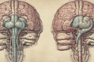
Introduction to Cerebrum
- Largest part of the brain, located in anterior and middle cranial fossae.
- Composed of diencephalon (central core) and telencephalon (cerebral hemispheres).
- Divided by the longitudinal cerebral fissure, containing the falx cerebri and anterior cerebral arteries.
- Corpus callosum connects the two hemispheres across the fissure.
Poles of the Cerebrum
- Three distinct poles:
- Frontal pole (anterior)
- Occipital pole (posterior)
- Temporal pole (between frontal and occipital, angled downwards)
Borders and Surfaces of the Cerebrum
- Each hemisphere has three borders:
- Superomedial
- Inferolateral
- Inferomedial, with anterior (medial orbital border) and posterior (medial occipital border) subdivisions.
- Surfaces divided into:
- Superolateral surface
- Medial surface
- Inferior surface (further divided into anterior orbital part and posterior tentorial part).
Lobes of the Cerebrum
- Defined by prominent sulci:
- Central sulcus separates frontal and parietal lobes.
- Posterior ramus of the lateral sulcus aids in lobe delineation.
- Frontal lobe lies anterior to central sulcus.
- Parietal lobe located behind central sulcus.
- Occipital lobe is posterior to the first imaginary line connecting parieto-occipital sulcus and preoccipital notch.
- Temporal lobe is below the posterior ramus of the lateral sulcus.
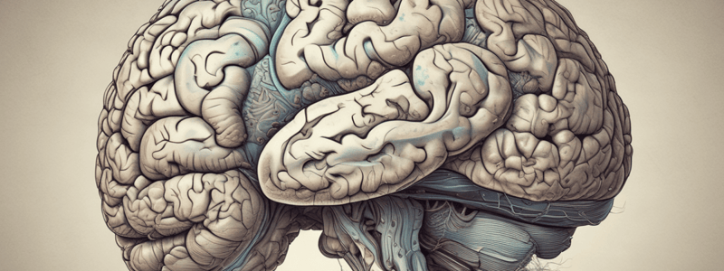
Gyrification
- Cerebral cortex exhibits gyri (ridges) and sulci (grooves) in most higher mammals, termed gyrencephalic brain.
- Lissencephalic brain observed in lower mammals; features smooth cortical surfaces.
Structures of the Frontal Lobe
- Precentral sulcus runs parallel to the central sulcus, with the area in between known as the precentral gyrus.
- Anteriorly, divided by superior and inferior frontal sulci into superior, middle, and inferior frontal gyri.
Structures of the Temporal Lobe
- Two parallel sulci, superior and inferior temporal sulci, divide the lobe into superior, middle, and inferior temporal gyri.
Structures of the Parietal Lobe
- Postcentral sulcus runs parallel to the central sulcus, with the area between them designated the postcentral gyrus.
- Divided into superior and inferior parietal lobules by the intraparietal sulcus.
Structures of the Occipital Lobe
- Contains short sulci, including lateral occipital sulcus, dividing it into superior and inferior occipital gyri.
Medial Surface Organizations
- Cingulate sulcus follows the contour of corpus callosum.
- Areas include gyri cinguli, paracentral lobule, medial frontal gyrus, and cuneus.
Inferior Surface Features
- Contains the olfactory sulcus and areas like gyrus rectus and medial/lateral occipitotemporal gyri.
Cerebral Cortex Functions
- Highest evolutionary development in humans.
- Composed of neurons, neuroglia, with approximately 14 billion neurons organized in layers.
- Functional roles include sensory perception, motor command execution, cognitive processes, and memory.
White Matter Classification
- Contains myelinated fibers categorized into association, commissural, and projection fibers.
- Association fibers interconnect areas within the same hemisphere, divided into short (local) and long (across lobes).
- Commissural fibers connect identical regions across hemispheres, with key examples including the corpus callosum and anterior commissure.
- Projection fibers extend from the cortex to subcortical centers, project both ways via corticofugal and corticopetal paths.
Functional Areas of the Cerebral Cortex
- Specific functions are attributed to different areas, classified according to sulci and gyri.
- Brodmann’s areas, numbering from 1 to 47, represent significant functional regions of the cerebral cortex.### Motor Area
- Located in the precentral gyrus on the superolateral surface and anterior paracentral lobule on the medial surface.
- Corresponds to Brodmann area 4 and possibly part of area 6 in the precentral gyrus.
- Specific regions control movements for different body parts:
- Lower limbs in the paracentral lobule
- Trunk and upper limbs in the upper part of the precentral gyrus
- Face and head in the lower part of the precentral gyrus.
Premotor Area
- Positioned anterior to the motor area, spanning the posterior parts of the superior, middle, and inferior frontal gyri.
- Corresponds to Brodmann areas 6 and 8 in superior and middle frontal gyri, areas 44 and 45 in inferior frontal gyrus (motor speech area of Broca).
- Stimulation leads to more intricate movements compared to the motor area.
- Closely related areas include:
- Motor speech area of Broca in inferior frontal gyrus.
- Frontal eye field.
Sensory Area
- Located in the postcentral gyrus, corresponds to Brodmann areas 1, 2, and 3.
- Extends to the medial hemisphere in the posterior portion of the paracentral lobule.
- Responses can be recorded based on individual body part stimulation.
- Represents different body parts, allowing for a mapped sensory area.
Visual Areas
- Found mostly in the occipital lobe on the medial surface, above and below the calcarine sulcus (area 17).
- Area 17 connects with areas 18 and 19, which mainly interpret visual impulses from area 17.
- Areas 18 and 19 are referred to as psychovisual areas.
Auditory Area
- Located in the temporal lobe, specifically the superior temporal gyrus.
- Contains the anterior transverse temporal gyrus (area 41) and extends slightly onto the superior temporal gyrus (areas 41, 42).
Applied Anatomy
- Lesions in Brodmann area 4 cause hemiplegia, evident as paralysis in the opposite body side.
- Lesions in the premotor area lead to apraxia, impacting skillful voluntary muscle movements despite normal function of the primary motor area.
- Stroke is defined as abrupt loss of brain function due to hemorrhage or inadequate blood supply, lasting over 24 hours.
Functional Deficits from Cerebral Damage
- Frontal lobe injuries may cause behavioral changes and problem-solving deficits.
- Parietal lobe damage often results in contralateral hemispatial neglect syndrome.
- Temporal lobe lesions can lead to auditory agnosia and prosopagnosia.
- Occipital lobe damage typically results in visual field defects, such as contralateral hemianopia or quadrantanopia with macular sparing.
- Global lesions may cause severe cognitive deficits (dementia), impairing the ability to answer basic questions.
Surfaces of the Cerebrum
- Divided into three main surfaces:
- Superolateral Surface
- Medial Surface
- Inferior Surface (subdivided into anterior orbital and posterior tentorial parts)
Lobes of the Cerebrum
- Boundaries of lobes are determined by prominent sulci and regions on each hemisphere.
- Superolateral Surface features two significant sulci:
- Posterior Ramus of the Lateral Sulcus: Starts near the temporal pole, curving slightly upwards to the back.
- Central Sulcus: Begins near the midpoint between the frontal and occipital poles, running downwards and forwards.
Gyri and Sulci
- Lissencephalic Brain: Refers to the smooth cerebral cortex in lower mammals, birds, and reptiles.
- Sulci vary in length and depth, separating adjacent gyri and lined by walls and a floor.
Frontal Lobe
- Precentral Sulcus: Runs parallel to the central sulcus, located a little anterior to it.
- Area between the precentral and central sulci is known as the Precentral Gyrus.
- Superior and inferior frontal sulci divide this region into superior, middle, and inferior frontal gyri.
- Anterior and ascending rami of the lateral sulcus extend into the inferior frontal gyrus, further dividing it.
Temporal Lobe
- Features Superior and Inferior Temporal Sulci parallel to the posterior ramus of the lateral sulcus.
- These sulci separate the superolateral surface into superior, middle, and inferior temporal gyri.
Parietal Lobe
- Postcentral Sulcus: Runs downwards and forwards, parallel to the central sulcus.
- The area between the postcentral and central sulci is named the Postcentral Gyrus.
Basal Ganglia and Surrounding Structures
- Corpus Striatum: Comprised of the caudate nucleus and lentiform nucleus, derived from the telencephalon.
- Claustrum: A thin layer of grey matter located between the lentiform nucleus and the insula.
- Internal Capsule: Contains major ascending and descending tracts, situated between the thalamus and the caudate nucleus.
White Matter of the Cerebrum
- Composed of myelinated nerve fibers (different diameters) and neuroglial cells.
- Forms the medullary substance beneath the cortical grey matter.
- Corona Radiata: White matter that radiates from the upper end of the internal capsule to the cortex.
Classification of White Matter
-
Divided into three groups:
- Association Fibers: Interconnect different areas within the same hemisphere without crossing the midline.
- Commissural Fibers: Connect corresponding areas between the two hemispheres.
- Projection Fibers: Extend between the cortex and subcortical structures.
-
No content or information was provided for summarization.
-
Please provide text or specific questions to generate study notes.
Studying That Suits You
Use AI to generate personalized quizzes and flashcards to suit your learning preferences.