Podcast
Questions and Answers
What is the purpose of tissue fixation during the preparation of histopathological specimens?
What is the purpose of tissue fixation during the preparation of histopathological specimens?
Which step in tissue processing involves cutting the specimen into ultra-thin sections for microscopic examination?
Which step in tissue processing involves cutting the specimen into ultra-thin sections for microscopic examination?
What is the main reason for using different staining techniques in histopathology?
What is the main reason for using different staining techniques in histopathology?
During which stage of intra-laboratory preparation is tissue dehydrated, cleared, and embedded?
During which stage of intra-laboratory preparation is tissue dehydrated, cleared, and embedded?
Signup and view all the answers
What is essential for ensuring the quality control of pathology specimens during both pre-laboratory and intra-laboratory phases?
What is essential for ensuring the quality control of pathology specimens during both pre-laboratory and intra-laboratory phases?
Signup and view all the answers
What is the main purpose of hematoxylin in H&E staining?
What is the main purpose of hematoxylin in H&E staining?
Signup and view all the answers
What does Masson's Trichrome staining differentiate between?
What does Masson's Trichrome staining differentiate between?
Signup and view all the answers
Which step involves applying dyes to enhance the visibility of tissue structures?
Which step involves applying dyes to enhance the visibility of tissue structures?
Signup and view all the answers
What is the purpose of periodic acid-Schiff (PAS) staining?
What is the purpose of periodic acid-Schiff (PAS) staining?
Signup and view all the answers
Which of the following is NOT a part of the key histopathological steps?
Which of the following is NOT a part of the key histopathological steps?
Signup and view all the answers
What additional diagnostic information can immunohistochemistry (IHC) provide?
What additional diagnostic information can immunohistochemistry (IHC) provide?
Signup and view all the answers
Which step in histopathology involves stabilizing and preserving tissue after collection?
Which step in histopathology involves stabilizing and preserving tissue after collection?
Signup and view all the answers
Why is staining considered critical in histopathological analysis?
Why is staining considered critical in histopathological analysis?
Signup and view all the answers
What is the primary purpose of fixation in biopsy preparation?
What is the primary purpose of fixation in biopsy preparation?
Signup and view all the answers
What is the significance of decalcification in the preparation of bone samples?
What is the significance of decalcification in the preparation of bone samples?
Signup and view all the answers
During the processing of biopsies, which step occurs immediately after fixation?
During the processing of biopsies, which step occurs immediately after fixation?
Signup and view all the answers
Which staining technique is commonly used after sectioning tissues for microscopic examination?
Which staining technique is commonly used after sectioning tissues for microscopic examination?
Signup and view all the answers
What is a critical factor to ensure accuracy in diagnosing tissues?
What is a critical factor to ensure accuracy in diagnosing tissues?
Signup and view all the answers
Which of the following statements best describes the processing of bone and biopsy tissues?
Which of the following statements best describes the processing of bone and biopsy tissues?
Signup and view all the answers
What purpose does sectioning serve in the examination of tissues?
What purpose does sectioning serve in the examination of tissues?
Signup and view all the answers
Why is it important to handle biopsies carefully during preparation?
Why is it important to handle biopsies carefully during preparation?
Signup and view all the answers
Study Notes
Pre-laboratory Specimen Handling
- Samples are collected via surgical procedures or autopsies.
- Specimens require proper identification, labelling, and documentation.
- Tissues are placed in an appropriate medium or fixative to prevent degradation
Intra-laboratory Tissue Handling and Preparation
- Trimming: Tissue is trimmed to the appropriate size for processing.
- Fixation: Tissue is stabilized to prevent decomposition through a process called autolysis and putrefaction.
- Processing: Tissue undergoes dehydration, clearing, and embedding stages.
- Embedding: Tissue is embedded in a solid medium (paraffin wax) to facilitate slicing into thin sections.
- Sectioning: Extremely thin slices are cut (around 3 to 5 micrometers thick) for examination with a microscope.
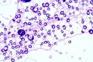
- Staining: Tissues are stained to highlight specific cell structures or components.
Common Staining Techniques
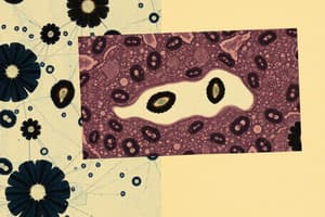
- Hematoxylin and Eosin (H&E Staining): Hematoxylin stains the nuclei blue/purple, while Eosin stains the cytoplasm and extracellular matrix pink/red. This is used to visualize general tissue morphology.
- Periodic Acid-Schiff (PAS Staining): Stains carbohydrates, highlighting basement membranes, fungal organisms, and glycogen storage diseases
- Masson's Trichrome: Differentiates muscle fibers (red), collagen (blue/green), and nuclei (black), mainly used for liver fibrosis.
- Immunohistochemistry (IHC): Uses antibodies to target specific antigens in the tissue to identify specific cell types, proteins, and receptors for cancer diagnosis and subtyping.
Summary of Key Histopathological Steps
- Specimen Collection: Proper handling and labeling.
- Fixation: Stabilization and preservation of tissue.
- Processing: Dehydration, clearing, and embedding of the specimen.
- Sectioning: Thin slices are cut for microscopic examination.
- Staining: Application of dyes to highlight tissue structures.
- Examination: Analysis under a microscope for diagnosis.
Bone Decalcification
- Bone requires decalcification for sectioning, as it is too hard to cut.
- Decalcified bone is then embedded in paraffin wax for sectioning.
- Decalcification is achieved by using acids to dissolve the calcium salts in the bone.
Biopsy Preparation
- Biopsies are small tissue samples taken from suspicious areas.
- Biopsies must be handled carefully to maintain their structure.
- Fixation: Biopsies are placed in a fixative (like formalin) immediately after removal to prevent degradation.
- Processing: Biopsies undergo the same processing steps as other tissues (dehydration, clearing, and embedding).
- Sectioning: Embedded biopsy can be sliced into thin sections for microscopic examination.
Special Considerations
- Orientation or positioning: Both bone and biopsy samples require careful positioning for embedding to ensure informative areas are ready for examination under the microscope.
- Staining: Sectioned bone and biopsy samples often require staining (H&E) for visualization of cellular structures to help identify abnormalities.
Key Points
- Decalcification is essential for bone sectioning to soften it so it can be sliced.
- Biopsies require rapid fixation to preserve cellular details.
- Both bone and biopsy tissues are processed similarly, but require specific handling techniques to ensure accurate diagnosis.
Studying That Suits You
Use AI to generate personalized quizzes and flashcards to suit your learning preferences.
Related Documents
Description
This quiz covers essential techniques for specimen handling and preparation in histology, including proper identification, fixation, and staining methods. Learn the steps involved in trimming, embedding, and sectioning tissue for microscopic examination. Test your knowledge on common staining techniques such as H&E staining.