36 Questions
What is the function of the fibrous skeleton of the heart?
To limit the spread of action potentials to specific pathways
Which of the following chambers of the heart receives oxygenated blood from the pulmonary veins?
Left atrium
What is the purpose of the coronary circulation?
To provide a functional blood supply to the heart muscle itself
What is the function of the chordae tendineae in the heart?
To support the valve flaps during ventricle contraction
Which of the following is NOT a part of the intrinsic cardiac conduction system?
Medulla oblongata
What is the sequence of blood flow through the heart?
Right atrium → tricuspid valve → right ventricle → pulmonary semilunar valve → pulmonary trunk → lungs → pulmonary veins → left atrium
What is the primary function of the atrioventricular (AV) node in the heart?
To delay impulses approximately 0.1 second
What is the term for the composite of all action potentials generated by nodal and contractile cells at a given time?
Electrocardiogram (ECG or EKG)
Which part of the brain is responsible for modifying the heartbeat through the autonomic nervous system?
Medulla oblongata
What is the name of the pathway that carries impulses from the atrioventricular (AV) bundle to the apex of the heart?
Purkinje fibers
What is the purpose of the cardiovascular center in the medulla oblongata?
To modify the heartbeat through the autonomic nervous system
What is the term for the potentials generated by autorhythmic cells due to slow open Na⁺ channels?
Pacemaker potentials
What is the name of the phase of the cardiac cycle where the ventricles relax and fill with blood?
Ventricular filling
What type of sounds are associated with the closing of heart valves?
Lub-dup
What is the name of the electrical event that represents ventricular repolarization?
T wave
What percentage of blood flows into the ventricles during ventricular filling?
80%
What is the name of the phase of the cardiac cycle where the atria relax and the ventricles begin to contract?
Ventricular systole
What is the term for the volume of blood remaining in each ventricle at the end of ventricular systole?
End systolic volume
What is the primary event that occurs during the isovolumetric contraction phase of the cardiac cycle?
All valves are open
During which phase of the cardiac cycle does the atrial systole occur?
Mid-to-late diastole
What is the term for the volume of blood in each ventricle at the end of ventricular diastole?
End diastolic volume
Which event is associated with the 'Lub' sound of the heart?
Closure of atrioventricular valves
What is the electrical event that represents ventricular depolarization?
QRS complex
What is the percentage of blood that flows into the ventricles during ventricular filling?
80%
What is the primary function of the myocardium layer of the heart wall?
To anchor cardiac muscle fibers and support great vessels and valves
What is the purpose of the spiral bundles of cardiac muscle cells in the myocardium?
To generate a coordinated contraction of the heart chambers
Which heart valve prevents backflow into the ventricles when they relax?
Pulmonary semilunar valve
What is the pathway of blood flow through the heart during the pulmonary circuit?
Right atrium → tricuspid valve → right ventricle → pulmonary trunk → lungs
What is the term for the electrical events that occur in the heart and are responsible for coordinating the heartbeat?
Sequence of excitation
Which of the following is responsible for modifying the heartbeat through the autonomic nervous system?
Cardiovascular center
What is the primary function of the purkinje fibers in the heart?
To complete the pathway into the apex and ventricular walls
What is the purpose of the cardioinhibitory center in the heart?
To decrease the heart rate through parasympathetic fibers
What is the term for the potentials generated by autorhythmic cells due to the opening of slow Na⁺ channels?
Pacemaker potentials
What is the sequence of electrical events in the heart?
SA node, AV node, AV bundle, Purkinje fibers
What is the role of the atrioventricular bundle in the heart?
To provide the only electrical connection between the atria and ventricles
What is the purpose of the cardioacceleratory center in the heart?
To increase the heart rate through sympathetic neurons
Study Notes
The Heart
- The heart is located in the thoracic cavity, surrounded by the pericardium.
- The heart wall consists of three layers: epicardium, myocardium, and endocardium.
Layers of the Heart Wall
- Epicardium: the visceral layer of the serous pericardium.
- Myocardium:
- Consists of spiral bundles of cardiac muscle cells.
- Has a fibrous skeleton that limits the spread of action potentials to specific pathways, anchors cardiac muscle fibers, and supports great vessels and valves.
- Endocardium: continuous with the lining of blood vessels, composed of simple squamous epithelium.
Heart Chambers
- Atria: the receiving chambers, with walls ridged by pectinate muscles.
- Right atrium receives deoxygenated blood from the superior and inferior vena cava, and coronary sinus.
- Left atrium receives oxygenated blood from the right and left pulmonary veins.
- Ventricles: the discharging chambers, with walls ridged by trabeculae carneae.
- Make up the largest part of the heart.
- Deoxygenated blood leaving the right ventricle enters the pulmonary trunk (to the lungs).
- Oxygenated blood leaving the left ventricle enters the aorta (to the body).
Heart Pathway of Blood Through the Heart
- The heart is two side-by-side pumps:
- Right side pumps blood to and from the lungs (pulmonary circuit).
- Left side pumps blood to and from all body tissue (systemic circuit).
- Pathway of blood through the heart:
- Right atrium → tricuspid valve → right ventricle → pulmonary semilunar valve → pulmonary trunk → pulmonary arteries → lungs.
- Lungs → pulmonary veins → left atrium → bicuspid valve → left ventricle → aortic semilunar valve → aorta → systemic circulation.
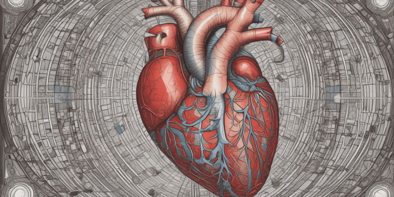
Coronary Circulation
- The functional blood supply to the heart muscle itself.
- Coronary arteries:
- Right and left coronary arteries (in atrioventricular groove).
- Marginal, circumflex, and anterior interventricular arteries.
- Coronary veins:
- Small cardiac, anterior cardiac, and great cardiac veins.
Heart Valves
- Ensure unidirectional blood flow through the heart.
- Atrioventricular (AV) valves:
- Prevent backflow into the atria when ventricles contract.
- Tricuspid valve (right) and bicuspid (Mitral) valve (left).
- Chordae tendineae support valve flaps during ventricle contraction.
- Flaps open during relaxation of the ventricles.
- Semilunar (SL) valves:
- Prevent backflow into the ventricles when ventricles relax.
- Aortic semilunar valve and pulmonary semilunar valve.
- Contraction increases pressure, forcing these valves open.
- Relaxation decreases pressure, causing these valves to close.
Heart Physiology: Electrical Events
- Intrinsic cardiac conduction system:
- A network of noncontractile (autorhythmic) cells that initiate and distribute impulses to coordinate the depolarization and contraction of the heart.
- Intrinsic cardiac conduction system:
- Sinoatrial (SA) node (pacemaker) generates impulses about 75 times/minute.
- Atrioventricular (AV) node delays impulses approximately 0.1 second.
- Atrioventricular (AV) bundle (bundle of His) is the only electrical connection between the atria and ventricles.
- Right and left bundle branches are the two pathways in the interventricular septum that carry the impulses toward the apex of the heart.
- Purkinje fibers complete the pathway into the apex and ventricular walls.
Heart Physiology: Sequence of Excitation
-
- P wave: depolarization of the SA node.
-
- QRS complex: ventricular depolarization.
-
- T wave: ventricular repolarization.
Heart Sounds
- Two sounds (lub-dup) associated with the closing of heart valves.
- "Lub" is the first sound, occurring when AV valves close, signifying the beginning of ventricular systole.
- "Dup" is the second sound, occurring when SL valves close at the beginning of ventricular diastole.
- Heart murmurs are abnormal heart sounds, often indicative of valve problems.
Mechanical Events: The Cardiac Cycle
- The cardiac cycle is all events associated with blood flow through the heart during one complete heartbeat.
- Phases of the cardiac cycle:
- Ventricular filling: takes place in mid-to-late diastole, with AV valves open, and 80% of blood passively flowing into ventricles.
- Atrial systole occurs, delivering the remaining 20% of blood.
- End diastolic volume (EDV) is the volume of blood in each ventricle at the end of ventricular diastole.
- Ventricular systole: atria relax and ventricles begin to contract.
- Rising ventricular pressure results in the closing of AV valves.
- Isovolumetric contraction phase (all valves are closed).
- In the ejection phase, ventricular pressure exceeds pressure in the large arteries, forcing the SL valves open.
- End systolic volume (ESV) is the volume of blood remaining in each ventricle.
Learn about the structure of the heart, including its location, layers, and functions. This quiz covers the epicardium, myocardium, endocardium, and more. Test your knowledge of the heart's anatomy and physiology
Make Your Own Quizzes and Flashcards
Convert your notes into interactive study material.
Get started for free