183 Questions
What type of joints are formed by the articular processes of vertebrae?
Zygapophysial (facet) joints
Which region of the vertebral column has the greatest range and variety of movement?
Cervical region
What is the characteristic of the vertebral body in cervical vertebrae?
Small and wider from side to side than anteroposteriorly
What is the distinctive feature of each cervical vertebra?
The transverse foramen in the transverse process
Which cervical vertebra is known as 'vertebra prominens'?
C7
What is the characteristic of the articular processes in cervical vertebrae?
Superior facets directed superoposteriorly
What is the characteristic of the vertebral arches in C1 (Atlas)?
Two lateral masses connected by anterior and posterior arches
Why is C1 (Atlas) named after Greek mythology?
Because it bears the weight of the cranium on its shoulders
Which part of the sacrum articulates with the coccyx?
Apex of Sacrum
What is the name of the opening at the inferior end of the dorsal sacrum?
Sacral Hiatus
How many small vertebrae typically fuse together to form the coccyx in adults?
4-5
What is the name of the joint formed by the base of the coccyx and the apex of the sacrum?
Sacrococcygeal Symphysis
At which week of embryonic development does ossification of vertebrae begin?
8th week
What is the name of the primary ossification center that will become the body of the vertebra?
Endrochondral Centrum
What is the name of the bony parts that unite with the vertebral body by age 25?
Secondary Ossification Centers
Which vertebra is an exception and lacks a centrum?
C1
What type of joint is the atlanto-occipital joint?
Synovial joint
Which movement is not permitted by the atlanto-occipital joints?
Circumduction
What is the main function of the anterior and posterior atlanto-occipital membranes?
To prevent excess movement of the atlanto-occipital joints
What type of joint is the median atlanto-axial joint?
Pivot joint
What is the function of the transverse ligament of the atlas?
To hold the dens in place against the atlas
What prevents excess rotation of the atlanto-axial joints?
The alar ligaments
What type of movement is characterized by flexion and extension in a frontal plane?
Lateral flexion and extension
Which region of the vertebral column has the most lateral flexion and extension?
Cervical region
What is the unique feature of the Cranial Shift?
Cervical rib articulating with C7 and a diminished 12th rib
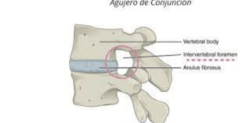
What is the main function of Intervertebral (IV) Discs?
Serve as shock absorbers and provide strong attachments between the vertebral bodies
What is the composition of the Anulus Fibrous?
Concentric lamellae
Where is the Nucleus Pulposus positioned within the disc?
Between the center and posterior aspect of the disc
What is unique about the L5-S1 disc?
It is the most inferior functional disc
What is the function of the Uncovertebral Joints?
To cause neck pain or bone spurs in later years
Where are the Uncovertebral Joints commonly found?
Between C3-C4 or C6-C7
How does the Nucleus Pulposus receive nourishment?
Through diffusion from blood vessels at the periphery of the anulus fibrosus and vertebral body
Where do flexion and extension movements primarily occur?
Cervical and Lumbar regions
What type of curvatures are the Thoracic and Sacral Kyphoses?
Primary Curvatures
What is the purpose of the curvatures of the vertebral column?
To provide resilience, flexibility, and shock-absorbance to the axial skeleton
What is unique about the Cervical lordosis in females?
It is more pronounced in females
What is the function of the spinal branches of the major cervical and segmental arteries?
To supply the vertebral column
What is the purpose of the internal and external vertebral venous plexuses?
To collect blood from the vertebrae
What is the function of the basivertebral veins?
All of the above
What is the function of the recurrent meningeal branches of the spinal nerves?
To supply most bone, IV discs, and ligaments as well as the meninges of the spinal cord
What is the function of the zygapophysial joints?
To be innervated by medial branches of adjacent posterior rami
What is the function of the intervertebral veins?
To receive veins from the spinal cord and vertebral venous plexuses
What is the primary attachment site of the Longissimus muscle?
Iliac crest and sacrum
What is the function of the Longissimus muscle?
It assists in extension of the vertebral column
Where do the fibers of the Longissimus muscle run?
Superiorly to the posterior surface of the ribs
What is the action of the Longissimus muscle when it contracts bilaterally?
It causes extension of the vertebral column
What is the action of the Longissimus muscle when it contracts unilaterally?
It causes lateral flexion of the head
What is the attachment site of the Longissimus muscle in the cervical region?
Mastoid process of the temporal bone
Where do the fibers of Multifidus pass?
Obliquely superomedially to Posterior rami of spinal nerves
From which anatomical structure does Multifidus arise?
Aponeurosis of erector spinae and sacroiliac ligaments
What is the primary function of Multifidus?
Stabilization of vertebrae during local movements
In which region is Multifidus thickest?
Lumbar region
What is the attachment site of Multifidus?
Spinous processes, entire length
Which of the following is NOT an origin of Multifidus?
Spinous process of T12
Which muscle lies deep to the trapezius, sternocleidomastoid, splenius, and semispinalis muscles?
Suboccipital muscles
Which nerve emerges as the vertebral artery courses deeply between the occipital bone and the atlas?
Posterior ramus of C1
What is the main function of the obliquus capitis inferior muscle?
Rotating the head on vertebra C1 and C2
What is the superior boundary of the suboccipital triangle?
Rectus capitis posterior major muscle
Which muscle is responsible for the lateral flexion of the head?
Longus capitis muscle
What is the function of the vertebral artery?
Supplying the spinal cord
What is the main function of the spinal cord?
Acting as a reflex center and conduction pathway
What is the characteristic of the spinal cord?
It is a continuation of the medulla oblongata
What is the function of the conus medullaris?
Forming the cauda equina
What is the characteristic of the cauda equina?
It is a bundle of spinal nerve roots
Which muscle forms the longitudinal bulge in the back of the neck near the median plane?
Semispinalis capitis
Which muscle is the middle layer of the transversospinalis muscle group?
Multifidus
Which muscle aids in lateral flexion of the vertebral column and stabilizes the vertebral column when acting bilaterally?
Intertransversarii
Which muscle elevates the ribs and assists with respiration?
Levatores costarum
Which muscle group originates from the transverse processes of vertebrae and passes to the spinous processes of more superior vertebrae?
Transversospinalis
Which muscle has fibers that run superomedially to the occipital bone and spinous processes in the thoracic and cervical regions?
Semispinalis
Which muscle group is responsible for extension of the vertebral column?
Transversospinalis
Which muscle is the deepest of the three layers in the transversospinalis muscle group?
Rotatores
Which muscle has fibers that run superiorly to the cranium?
Spinalis
Which muscle group consists of semispinalis, multifidus, and rotatores muscles?
Transversospinalis
What is the course of the anterior spinal artery?
Runs inferiorly in the anterior median fissure
What is the function of the sulcal arteries?
Supply approximately two thirds of the cross-section of the spinal cord
What is the origin of the posterior spinal arteries?
Branches of the posterior inferior cerebellar artery
What is the function of the segmental medullary arteries?
Supply much of the spinal cord, including the lumbosacral enlargement
Where do the segmental medullary arteries occur mainly?
In association with the cervical and lumbosacral enlargements
What is the great anterior segmental medullary artery?
A large artery that reinforces the circulation to two thirds of the spinal cord
What is the function of the radicular arteries?
Supply the nerve roots and their coverings
What occurs at the irregular levels where the radicular arteries occur?
The segmental medullary arteries replace the radicular arteries
What is the shape of the thoracic cavity?
Truncated cone
What is the function of the thoracic cage?
To protect vital thoracic and abdominal organs from external forces
What is the floor of the thoracic cavity?
The diaphragm
What is the primary function of the thoracic wall?
To protect the thoracic and abdominal organs
What is the composition of the thoracic skeleton?
Ribs, thoracic vertebrae, costal cartilages, and intervertebral discs
What is the characteristic of the thoracic cavity's central compartment?
It contains the thoracic viscera except for the lungs
What is the function of the thoracic cage in relation to the upper limbs?
To support the weight of the upper limbs
What type of bones are the ribs?
Flat bones
What is the primary function of the diaphragm?
To facilitate breathing by increasing the volume of the thoracic cavity
What is the significance of the pleural reflections and recesses?
They facilitate the movement of the lungs during breathing
What is the function of the intercostal muscles?
To facilitate movement of the ribs during breathing
What is the characteristic of the visceral pleura?
It is a layer of serous membrane surrounding the lungs
What is the function of the thoracic wall vasculature?
To supply oxygenated blood to the thoracic muscles
What is the significance of the bony anatomy of the thorax?
It protects vital organs in the thoracic cavity
Lymph from the inferior quadrants of the breast may pass deeply to which lymph nodes?
Abdominal lymph nodes
The clavicles demarcate the superior division between which zones of lymphatic drainage?
Above clavicles: inferior jugular lymph nodes; below clavicles: axillary lymph nodes
The anterior median line (AML) indicates the intersection of the median plane with which part of the thoracic wall?
Anterior thoracic wall
The midclavicular line (MCL) passes through which part of the clavicle?
Midpoint of the clavicle
The posterior axillary line (PAL) is drawn vertically along which anatomical structure?
Posterior axillary fold
The posterior median line (PML) is a vertical line along which anatomical structure?
Tips of the spinous processes of the vertebrae
Which anatomical structure forms the anterior axillary fold?
Pectoralis major and teres major muscles
The sternal angle is located at the level of which costal cartilages?
2nd pair of costal cartilages
What type of blood does the pulmonary veins carry?
Oxygen-rich blood
What is the main function of the parasympathetic fibers in the pulmonary plexus?
To stimulate the glands of the bronchial tree
What is the origin of the bronchial arteries?
Aorta
What is the primary function of the sympathetic fibers in the pulmonary plexus?
To inhibit alveolar glands
What is the function of the visceral afferent fibers in the pulmonary plexus?
To transmit pain impulses
What is the superficial subpleural lymphatic plexus responsible for?
Draining the lung parenchyma
Where do the lymphatic vessels from the superficial subpleural lymphatic plexus drain into?
Bronchopulmonary lymph nodes
What is the primary function of the nerves of the lungs and pleura?
To transmit sensory information
Which part of the diaphragm arises from the anterior surfaces of the bodies of the superior three lumbar vertebrae?
Crura
What is the formation of the esophageal hiatus?
Right crus
What attaches to the internal surfaces of the inferior six costal cartilages and adjoining ribs on each side?
Costal part
What attaches to the posterior aspect of the xiphoid process?
Sternal part
What forms the aortic hiatus?
Fibrous medial arcuate ligament and crura
What attaches to the medial and lateral arcuate ligaments on each side?
Diaphragm
What is formed by the right and left muscular crura that ascend to the central tendon?
Right and left domes
What arises from the anterior longitudinal ligament and the IV discs?
Crura
Where is the Sinoatrial Node located?
In the wall of the left atrium near the opening of the superior vena cava
What structure receives the great cardiac vein at its left end?
Coronary sinus
Which vein opens into the coronary sinus?
Left posterior ventricular vein
What is the function of the cardiac conduction system?
To generate and transmit impulses that produce coordinated contractions of the heart
Where is the Atrioventricular Bundle located?
In the upper part of the interventricular septum
What is the purpose of the cardiac conduction system?
To generate and transmit impulses that produce coordinated contractions of the heart
What forms the base of the heart?
Left and right atria
Which surface of the heart is formed mainly by the right atrium?
Right pulmonary surface
Which border of the heart is formed mainly by the right atrium?
Right border
What receives the pulmonary veins?
Left atrium
What is the relation of the base of the heart to the vertebral bodies?
It faces posteriorly toward the bodies of vertebrae T6–T9
What is the diaphragmatic surface of the heart formed mainly by?
Left ventricle
What is the superior border of the heart formed by?
Right and left atria and auricles
What is the left pulmonary surface of the heart formed mainly by?
Left ventricle
What is the mediastinum occupied by?
Mass of tissue between the two pulmonary cavities
What is the pericardium composed of?
Two layers: fibrous and serous
What is the inferior limit of the superior mediastinum?
Horizontal plane that includes the sternal angle
What is the middle mediastinum comprised of?
Pericardium and its contents only
What is the purpose of the mediastinal pleura?
To cover the mediastinum on each side
What is the thoracic duct located in?
Posterior mediastinum
What forms the right border of the heart?
Right atrium
What is the origin of the ascending aorta?
Aortic orifice of the left ventricle
What is the function of the ear-like right auricle?
To increase the capacity of the atrium
What separates the ridged muscular wall of the inflow part of the right ventricle from the smooth wall of the conus arteriosus?
Supraventricular crest
What is the course of the arch of aorta?
Arches posteriorly on the left side of the trachea and esophagus
What receives blood from the right atrium through the right AV orifice?
Right ventricle
What is the origin of the posterior intercostal arteries?
Thoracic descending aorta
What is the distribution of the vagus nerve (CN X)?
Pulmonary plexus, esophageal plexus, and cardiac plexus
What is the location of the right AV orifice?
Posterior to the body of the sternum at the level of the 4th and 5th intercostal spaces
What is the origin of the phrenic nerve?
Anterior rami of C3–C5 nerves
What is the characteristic of the interior of the right ventricle?
It has irregular muscular elevations (trabeculae carneae)
What is the course of the thoracic descending aorta?
Descends in the posterior mediastinum to the left of the vertebral column
What forms the largest part of the anterior surface of the heart?
Right ventricle
What is the origin of the bronchial arteries?
Anterior aspect of the thoracic aorta
What is the name of the arterial cone that the right ventricle tapers into superiorly?
Conus arteriosus
What is the distribution of the intercostal nerves?
Lower nerves supply muscles and skin of anterolateral abdominal wall
What is the origin of the esophageal arteries?
Anterior aspect of the thoracic aorta
What is the origin of the superior phrenic arteries?
Anterior aspect of the thoracic aorta
ما هو العضلات الأكثر احتمالاً لیدامجا أثناء استخدام الستيرويدات الابتنائية؟
العضلة المصطقة للفقرات الخلفية السفلى
ما هو Bereich الذي قد يواجه انخفا ضا في الحس بسبب ضغط العضلة المصطقة للفقرات الخلفية السفلى؟
الرقبة وعظم القبة
ما هو الأعراض الأكثر احتمالاً لمرضى الذين يعانون من صعوبة في الثني الجانبي للرقبة؟
إصابة العضلة المصطقة للفقرات الخلفية السفلى
ما هو موقع العضلة التي قد تتعرض للإصابة أثناء استخدام الستيرويدات الابتنائية؟
الرقبة
ما هو العلاج الأكثر احتمالاً لمرضى الذين يعانون من صعوبة في الثني الجانبي للرقبة؟
علاج العضلة المصطقة للفقرات الخلفية السفلى
ما هو النوع من الألم الذي يعاني منه المريض وفقاً للاستشارة الطبية؟
ألم حاد
ما هو الامتداد والحركة التي يتم السماح بها في المنطقة القطنية من العمود الفقري؟
اللateral flexion والاستدارة
ما هو المكون الرئيسي ل القرص بين الفقري؟
الأنulus الفيبروزي
ما هو الوسيط الرئيسي لتدفق الدم إلى النواة اللزجة؟
الشرايين الفقرية
ما هو الوسيط الرئيسي لحركة الوعاء في العمود الفقري؟
القرص بين الفقري
ما هو التشخيص في حالة الدراسة 2-11؟
إصابة/stretch في الرقبة
ما هو السبب الرئيسي لإصابة الرجل البالغ من العمر 45 سنة في الدراسة 2-11؟
حрупية Box من الكتب
ما هو الموقع الذي تت 发生的 الإصابة في الدراسة 2-11؟
المنطقة السفلية للعمود الفقري
ما هو النوع من الإصابة التي حدثت في الدراسة 2-11؟
إصابة بالمد والقوة
ما هو نوع الألم الذيperienced by the man in the case study 2-11؟
ألم شديد في المنطقة السفلية للعمود الفقري
أين يبدأ الألم في الساق في المثال المذكور؟
الضلع الوحشي للساق
ما هو الموقع الأولی ل xuất現 الألم في المثال؟
الضلع الوحشي للساق
ما هو الاتجاه الذي ينتقل فيه الألم في المثال؟
من الساق إلى القدم
ما هو الجزء الأخير الذي يصل إليه الألم في المثال؟
ال 腳
ما هو الجزء الأولي الذي يتم ملاحظة الألم فيه في المثال؟
الضلع الوحشي للساق
ما هو النتيجة الرئيسية لشلل مفصل المثانة والقناة الشرجية؟
شلل جزئي أو كلي في الساقين
ما هو الجزء الرئيسي للجسم الذي يتأثر به شلل مفصل المثانة والقناة الشرجية؟
الجهاز البولي
ما هو الاسم الطبي لشلل مفصل المثانة والقناة الشرجية؟
Paraplegia
ما هو النتيجة الثانية لشلل مفصل المثانة والقناة الشرجية؟
فقدان السيطرة على المثانة
ما هو التأثير الرئيسي لشلل مفصل المثانة والقناة الشرجية على الحياة اليومية؟
صعوبة في التحكم في المثانة
Study Notes
Regional Characteristics of Vertebrae
- Cervical vertebrae:
- Form the skeleton of the neck
- Smallest of the 24 movable vertebrae
- Located between the cranium and thoracic vertebrae
- Smaller size reflects the fact that they bear less weight than the larger inferior vertebrae
- Greatest range and variety of movement of all vertebral regions
- Distinctive features of cervical vertebrae:
- Vertebral body: small and wider from side to side than anteroposteriorly
- Superior surface concave with uncus of body (uncinate process)
- Inferior surface convex
- Transverse foramen: most distinctive feature of each cervical vertebra, found in the transverse process
- Articular processes: superior facets directed superoposteriorly; inferior facets directed infero-anteriorly
- Spinous process: short (C3-C5) and bifid (C3-C6); process of C6 long, that of C7 longer (vertebra prominens)
Atlanto-Axial Joints
- Formed between the atlas (C1) and axis (C2)
- Permit the head to be turned side to side (the "NO" movement)
- Cranial and C1 rotate on C2, with the dens as a pivot
- Three atlanto-axial articulations:
- Two lateral atlanto-axial joints: between interior facets of lateral masses of C1 and superior facets of C2
- One median atlanto-axial joint: between the dens (C2) and anterior arch of Atlas (C1)
Vertebral Column
- Four curvatures of the adult vertebral column: cervical, thoracic, lumbar, and sacral
- Primary curvatures: thoracic and sacral kyphoses, developed during the fetal period
- Secondary curvatures: cervical and lumbar lordoses, acquired in relation to the erect human posture
- Curvatures provide resilience, flexibility, and shock-absorbance to the axial skeleton
Vascularity and Nerve Supply of Vertebral Column
- Spinal branches of the major cervical and segmental arteries supply the vertebral column
- Venous drainage: internal and external vertebral venous plexuses collect blood from the vertebrae and drain into the vertebral veins of the neck and segmental veins of the trunk
- Nerve supply: recurrent meningeal branches of the spinal nerves supply most bone, IV discs, and ligaments as well as the meninges of the spinal cord
Ossification of Vertebrae
- Begins around the 8th week at the end of the embryonic period
- Three primary ossification centers develop in cartilaginous vertebrae:
- Endrochondral centrum
- Two perichondral centers in each half of the neural arch
- Ossification continues through fetal period
- At birth, vertebra consists of three bony parts united by hyaline cartilage
- Inferior sacral and coccygeal vertebrae still cartilaginous; ossify during infancy
Intrinsic Back Muscles
- The intrinsic back muscles consist of two groups: the major deep layer and the minor deep layer.
- The major deep layer includes the transversospinalis muscle group, which consists of semispinalis, multifidus, and rotatores.
- Semispinalis:
- Originates from transverse processes of vertebrae
- Passes to spinous processes of more superior vertebrae
- Divided into three parts: semispinalis capitis, semispinalis thoracis, and semispinalis cervicis
- Semispinalis capitis forms the longitudinal bulge in the back of the neck
- Multifidus:
- Originates from posterior sacrum, posterior superior iliac spine, and transverse processes of vertebrae
- Passes to spinous processes of more superior vertebrae
- Thickest in the lumbar region
- Stabilizes vertebrae during local movements
- Rotatores:
- Originates from transverse processes of vertebrae
- Passes to spinous processes of more superior vertebrae
- Best developed in the thoracic region
- Acts as stabilizers
- Semispinalis:
- The minor deep layer includes interspinales, intertransversarii, and levatores costarum.
- Interspinales:
- Aids in extension and rotation of vertebral column
- Intertransversarii:
- Aids in lateral flexion of vertebral column
- Acts bilaterally to stabilize vertebral column
- Levatores costarum:
- Elevates ribs and aids in respiration
- Assists in lateral flexion of vertebral column
- Interspinales:
Surface Anatomy of the Back
- The surface anatomy of the back includes the following landmarks:
- Nuchal groove
- Vertebra prominens (C7)
- Posterior median furrow
- Intergluteal cleft
- Erector spinae
- Trapezius
- Rhomboids
- Latissimus dorsi
- Median borders of scapulae
- Dimples overlying PSIS
- Site of sacrum
Suboccipital and Deep Neck Muscles
- The suboccipital region includes four small muscles:
- Rectus capitis posterior major
- Rectus capitis posterior minor
- Obliquus capitis superior
- Obliquus capitis inferior
- These muscles are mainly postural muscles, but they also act to produce movement of the head.
- The suboccipital muscles act on the head directly or indirectly by extending it on vertebra C1 and rotating it on C1 and C2.
Suboccipital Triangle
- The suboccipital triangle is bounded by:
- Superomedial boundary: posterior atlanto-occipital membrane
- Superolateral boundary: rectus capitis posterior major
- Inferolateral boundary: obliquus capitis inferior
- Floor: posterior arch of vertebra C1 (atlas)
- Roof: semispinalis capitis
- Contents: vertebral artery and suboccipital nerve
Principal Muscles Producing Movement of Atlanto-Occipital Joints
- Flexion:
- Longus capitis
- Rectus capitis anterior
- Anterior fibers of sternocleidomastoid
- Suprahyoid and infrahyoid muscles
- Extension:
- Rectus capitis posterior major and minor
- Obliquus capitis superior
- Longissimus capitis
- Splenius capitis
- Rotation:
- Obliquus capitis inferior
- Rectus capitis posterior major and minor
- Longissimus capitis
- Splenius capitis
Nerves of the Posterior Cervical Region
- The suboccipital nerve is a branch of the posterior ramus of spinal nerve C1.
- The greater occipital nerve is a branch of the posterior ramus of spinal nerve C2.
- The lesser occipital nerve is a branch of the anterior rami of spinal nerves C2-C3.
- The posterior rami of spinal nerves C3-C7 innervate the skin and muscles of the back and neck.
The Spinal Cord
-
The spinal cord is a major reflex center and conduction pathway between the body and brain.
-
It is protected by the vertebrae, their ligaments and muscles, the spinal meninges, and the cerebrospinal fluid.
-
The spinal cord occupies the superior two-thirds of the vertebral canal in adults.
-
It is a continuation of the medulla oblongata and extends from the foramen magnum in the occipital bone to the level of the L1 or L2 vertebra.
-
The spinal cord has two enlargements: the cervical enlargement and the lumbosacral enlargement.
-
The conus medullaris is the inferior, tapering end of the spinal cord, and it ends at the level of the L1 or L2 vertebra.
-
The cauda equina is the inferior continuation of the spinal nerve roots caudal to the termination of the spinal cord.### Arteries of Spinal Cord and Nerve Roots
-
The anterior spinal artery runs inferiorly in the anterior median fissure and is formed by the union of branches of the vertebral arteries.
-
Sulcal arteries arise from the anterior spinal artery and enter the spinal cord through the anterior median fissure, supplying approximately two-thirds of the cross-section.
Posterior Spinal Arteries
- Each posterior spinal artery is a branch of either the vertebral artery or the posteroinferior cerebellar artery.
- The posterior spinal arteries commonly form anastomosing channels in the pia mater.
Blood Supply to the Spinal Cord
- The anterior and posterior spinal arteries can supply only the short superior part of the spinal cord.
- Circulation to much of the spinal cord depends on segmental medullary and radicular arteries running along the spinal nerve roots.
Segmental Medullary Arteries
- The anterior and posterior segmental medullary arteries are derived from spinal branches of the ascending cervical, deep cervical, vertebral, posterior intercostal, and lumbar arteries.
- These arteries occur mainly in association with the cervical and lumbosacral enlargements, in regions where a good blood supply is needed.
The Great Anterior Segmental Medullary Artery
- The great anterior segmental medullary artery is on the left side in about 65% of people and reinforces the circulation to two-thirds of the spinal cord, including the lumbosacral enlargement.
- This artery, much larger than the other segmental medullary arteries, usually arises via a spinal branch from an inferior intercostal or upper lumbar artery and enters the vertebral canal through the IV foramen at the lower thoracic or upper lumbar level.
Radicular Arteries
- The posterior and anterior roots of the spinal nerves and their coverings are supplied by posterior and anterior radicular arteries, which run along the nerve roots.
- The radicular arteries do not reach the posterior, anterior, or spinal arteries, but are replaced by segmental medullary arteries at irregular levels.
###Thorax Anatomy
- The thorax, also known as the chest, is the part of the body between the neck and abdomen, broadest superiorly due to the presence of the pectoral girdle, pectoral and scapular muscles, and female breasts.
###Thoracic Cavity
- The thoracic cavity has the shape of a truncated cone, narrowest superiorly and increasing inferiorly.
- The thoracic cavity is divided into three major spaces: the central compartment or mediastinum (thoracic viscera except for lungs), right and left pulmonary cavities (housing the lungs).
- The floor of the thoracic cavity is the diaphragm.
###Thoracic Wall/Thoracic Cage
- The thoracic wall includes the thoracic cage, muscles that extend between the ribs, skin, subcutaneous tissue, muscles, and fascia covering the anterolateral aspect.
- Functions of the thoracic cage:
- Protect vital thoracic and abdominal organs from external forces.
- Resist the negative internal pressures generated by the elastic recoil of the lungs and inspiratory movements.
- Provide attachment for and support the weight of the upper limbs.
- Provide anchoring attachment of many muscles that move and maintain the position of the upper limbs relative to the trunk.
- Provide attachments for muscles of the abdomen, neck, back, and respiration.
###Thoracic Skeleton
- The thoracic skeleton includes 12 pairs of ribs and associated costal cartilages, 12 thoracic vertebrae, and intervertebral discs interposed between them, and the sternum.
###Ribs
- Ribs are curved, flat bones that form most of the thoracic cage.
- Each rib has a spongy interior containing bone marrow.
- There are three types of ribs.
###Surface Anatomy of the Thoracic Wall
- Clavicles lie subcutaneously, forming bony ridges at the junction of the thorax and neck.
- Clavicles demarcate the superior division between zones of lymphatic drainage (above clavicles inferior jugular lymph nodes; below the clavicles axillary lymph nodes).
- Sternum and jugular notch (at the inferior border of T2).
- Sternal angle: level of 2nd pair of costal cartilages.
###Vertebral Levels of Sternum and Transverse Thoracic Plane
- Anterior median (midsternal) line (AML) indicates the intersection of the median plane with the anterior thoracic wall.
- Midclavicular line (MCL) passes through the midpoint of the clavicle, parallel to the AML.
- Anterior axillary line (AAL) runs vertically along the anterior axillary fold formed by the inferolateral border of the pectoralis major as it spans from the thoracic cage to the humerus in the arm.
- Midaxillary line (MAL) runs from the apex (deepest part) of the axillary fossa (armpit), parallel to the AAL.
- Posterior axillary line (PAL), also parallel to the AAL, is drawn vertically along the posterior axillary fold formed by the latissimus dorsi and teres major muscles as they span from the back to the humerus.
- Posterior median (midvertebral) line (PML) is a vertical line along the tips of the spinous processes of the vertebrae.
###Lymphatic Drainage of the Thoracic Wall
- Most lymph from the medial breast quadrants drains to the parasternal lymph nodes or to the opposite breast, whereas lymph from the inferior quadrants may pass deeply to abdominal lymph nodes (subdiaphragmatic inferior phrenic lymph nodes).
###Lymphatic Drainage of the Lungs
- The pulmonary lymphatic plexuses communicate freely.
- The superficial subpleural lymphatic plexus lies deep to the visceral pleura and drains the lung parenchyma (tissue) and visceral pleura.
- Lymphatic vessels from this superficial plexus drain into the bronchopulmonary lymph nodes (hilar lymph nodes) in the region of the lung hilum.
###Nerves of the Lungs and Pleura
- Derived from the pulmonary plexuses anterior and mainly posterior to the roots of the lungs.
- Contain parasympathetic fibers:
- Motor to the smooth muscle of the bronchial tree (bronchoconstrictor).
- Inhibitory to the pulmonary vessels (vasodilator).
- Secretory to the glands of the bronchial tree (secretomotor).
- Sympathetic fibers:
- Inhibitory to the bronchial muscle (bronchodilator).
- Motor to the pulmonary vessels (vasoconstrictor).
- Inhibitory to the alveolar glands of the bronchial tree—type II secretory epithelial cells of the alveoli.
- Visceral afferent fibers:
- Reflexive (conducting subconscious sensations associated with reflexes that control function).
- Nociceptive (conducting pain impulses generated in response to painful or injurious stimuli, such as chemical irritants, ischemia, or excessive stretch).
###Diaphragm
- Crura of the diaphragm: musculotendinous bands that arise from the anterior surfaces of the bodies of the superior three lumbar vertebrae, the anterior longitudinal ligament, and the IV discs.
- Esophageal hiatus is a formation of the right crus.
- The right and left crura and the fibrous medial arcuate ligament form the Aortic hiatus.
- Diaphragm is also attached on each side to the medial and lateral arcuate ligaments.
The Mediastinum
- The mediastinum is the middle septum that occupies the space between the two pulmonary cavities and is the central compartment of the thoracic cavity.
- It is covered on each side by mediastinal pleura and contains all of the thoracic viscera except for the lungs.
- The mediastinum extends from the superior thoracic aperture to the diaphragm and from the sternum and costal cartilages to the thoracic vertebrae.
- It is a highly mobile area in living people.
Divisions of the Mediastinum
- The mediastinum is divided into superior and inferior parts.
- The superior mediastinum extends from the superior thoracic aperture to the horizontal plane that includes the sternal angle anteriorly and the junction of T4 and T5 vertebrae posteriorly.
- The inferior mediastinum is between the transverse thoracic plane and the diaphragm.
- The inferior mediastinum is further subdivided into anterior, middle, and posterior parts by the pericardium.
Pericardium
- The pericardium is a fibroserous membrane that covers the heart and the beginning of its great vessels.
- It is a closed sac composed of two layers: the fibrous pericardium and the serous pericardium.
- The fibrous pericardium is the tough, external layer, continuous with the central tendon of the diaphragm.
The Heart
- The heart is a muscular organ that pumps blood throughout the body.
- It has four chambers: the right atrium, left atrium, right ventricle, and left ventricle.
- The atria receive blood, while the ventricles pump blood out of the heart.
Surfaces of the Heart
- The anterior (sternocostal) surface of the heart is formed mainly by the right ventricle.
- The diaphragmatic (inferior) surface of the heart is formed mainly by the left ventricle and partly by the right ventricle.
- The right pulmonary surface of the heart is formed mainly by the right atrium.
- The left pulmonary surface of the heart is formed mainly by the left ventricle and forms the cardiac impression in the left lung.
Borders of the Heart
- The right border of the heart is formed by the right atrium and extends between the superior vena cava and the inferior vena cava.
- The inferior border of the heart is formed mainly by the right ventricle and slightly by the left ventricle.
- The left border of the heart is formed mainly by the left ventricle and slightly by the left auricle.
- The superior border of the heart is formed by the right and left atria and auricles.
Right Atrium
- The right atrium forms the right border of the heart and receives venous blood from the superior vena cava, inferior vena cava, and coronary sinus.
- The ear-like right auricle is a conical muscular pouch that projects from the chamber and increases the capacity of the atrium.
Interior of the Right Atrium
- The interior of the right atrium has a rough, trabeculated wall.
- The right atrium receives blood from the superior vena cava and inferior vena cava through the right atrioventricular orifice.
Right Ventricle
- The right ventricle forms the largest part of the anterior surface of the heart and small parts of the diaphragmatic and inferior surfaces.
- The right ventricle receives blood from the right atrium through the right atrioventricular orifice and pumps it into the pulmonary trunk.
Interior of the Right Ventricle
- The interior of the right ventricle has irregular muscular elevations (trabeculae carneae).
- A thick muscular ridge, the supraventricular crest, separates the ridged muscular wall of the inflow part of the chamber from the smooth wall of the outflow part.
Cardiac Conduction System
- The cardiac conduction system consists of nodal tissue that initiates and coordinates contractions of the four chambers.
- It generates and transmits impulses that produce the cardiac cycle.
- The cardiac conduction system includes the sinoatrial (SA) node, atrioventricular (AV) node, atrioventricular bundle (bundle of His), and right and left bundle branches.
Thoracic Aorta
- The thoracic aorta is the part of the aorta that lies in the thoracic cavity.
- It begins posterior to the 2nd right sternocostal joint at the level of the sternal angle and arches superiorly, posteriorly, and to the left.
- The thoracic aorta gives rise to several branches, including the brachiocephalic, left common carotid, and left subclavian arteries.
Branches of the Aorta
- The ascending aorta gives rise to the right and left coronary arteries.
- The arch of the aorta gives rise to the brachiocephalic, left common carotid, and left subclavian arteries.
- The thoracic aorta gives rise to posterior intercostal arteries, subcostal arteries, and some phrenic arteries.
Nerves of the Thorax
- The vagus nerve (CN X) enters the superior mediastinum posterior to the sternoclavicular joint and brachiocephalic vein and gives rise to the recurrent laryngeal nerve.
- The phrenic nerve arises from the anterior rami of C3-C5 nerves and passes through the superior thoracic aperture and runs between the mediastinal pleura and pericardium.
- The intercostal nerves (1-11) arise from the anterior rami of T1-T11 nerves and run in the intercostal spaces between the internal and innermost layers of intercostal muscles.
Muscle Damage and Hypertrophy
- External oblique muscle is most likely to be damaged.
- Anabolic steroid user may experience hypertrophy of the semispinalis capitis muscle.
Compression and Sensation
- Compression by the semispinalis capitis muscle can lead to reduced sensation in the scalp just posterior to the external ear.
Neck Flexion and Injury
- Difficulty in lateral flexion of the neck can be caused by hyperextension injury of the neck.
- Hyperextension injury of the neck can lead to acute back pain.
Case Study 2-11
- Sudden severe pain in the lower back can be caused by carrying a heavy load.
- Severe pain in the lower back can lead to a dull ache in the posterolateral aspect of the thigh, extending along the calf of the leg into the foot.
- Diagnosis of paraplegia and other neurological deficits can result in sphincter paralysis in the urinary bladder and anal canal.
This quiz covers the regional characteristics of cervical vertebrae, including their size, location, and range of movement. It also discusses their function and comparison to other vertebrae.
Make Your Own Quizzes and Flashcards
Convert your notes into interactive study material.
Get started for free