Podcast
Questions and Answers
What is the term for the joints between each bony rib and its respective costal cartilage, and what type of joint are they classified as?
What is the term for the joints between each bony rib and its respective costal cartilage, and what type of joint are they classified as?
Costochondral joints, synchondroses
What is the difference between the first sternocostal joint and the sternocostal joints between the sternum and the costal cartilage of ribs 2-7?
What is the difference between the first sternocostal joint and the sternocostal joints between the sternum and the costal cartilage of ribs 2-7?
The first sternocostal joint is a synchondrosis, while the sternocostal joints between the sternum and the costal cartilage of ribs 2-7 are synovial joints
What is the cause of costochondritis, and how is it typically treated?
What is the cause of costochondritis, and how is it typically treated?
The cause of costochondritis is usually unknown, but it can be caused by repeated minor trauma or infection; it is typically treated with NSAIDs and rest
What is the main function of the fibrocartilage in a symphysis?
What is the main function of the fibrocartilage in a symphysis?
What is an example of a symphysis, and what is its function in pregnant females?
What is an example of a symphysis, and what is its function in pregnant females?
What is the functional category that a symphysis is placed in, and why is it in this category?
What is the functional category that a symphysis is placed in, and why is it in this category?
What is the significance of intervertebral discs in the spine, and how do they contribute to the flexibility of the spine?
What is the significance of intervertebral discs in the spine, and how do they contribute to the flexibility of the spine?
How do the collective movements of intervertebral discs contribute to the flexibility of the spine?
How do the collective movements of intervertebral discs contribute to the flexibility of the spine?
Explain why the presence of a synchondrosis in a long bone indicates that the bone is still growing.
Explain why the presence of a synchondrosis in a long bone indicates that the bone is still growing.
What is the functional classification of a synchondrosis and why is this classification appropriate?
What is the functional classification of a synchondrosis and why is this classification appropriate?
Describe the process of how a synchondrosis becomes a synostosis, and provide an example of this process.
Describe the process of how a synchondrosis becomes a synostosis, and provide an example of this process.
Compare and contrast the structure and function of synchondroses and symphyses. Provide an example of each.
Compare and contrast the structure and function of synchondroses and symphyses. Provide an example of each.
Explain why the spheno-occipital synchondrosis is useful for determining the age of a skull.
Explain why the spheno-occipital synchondrosis is useful for determining the age of a skull.
Why is the immobility of synchondroses an important factor in the skeletal system?
Why is the immobility of synchondroses an important factor in the skeletal system?
Explain how the structure of hyaline cartilage in a synchondrosis contributes to its function.
Explain how the structure of hyaline cartilage in a synchondrosis contributes to its function.
Discuss the significance of synchondroses in the growth and development of the human skeleton.
Discuss the significance of synchondroses in the growth and development of the human skeleton.
Study Notes
Cartilaginous Joints
- Cartilaginous joints have cartilage between the articulating bones and lack a joint cavity.
- They can be either immobile or slightly mobile.
- The cartilage found between the articulating bones is either hyaline cartilage or fibrocartilage.
Synchondroses
- A synchondrosis is an articulation in which bones are joined by hyaline cartilage.
- Functionally, all synchondroses are immobile and thus classified as synarthroses.
- Examples of synchondroses include:
- Epiphyseal plates in children, which bind the epiphyses and diaphysis of long bones.
- The spheno-occipital synchondrosis, found between the body of the sphenoid and the basilar part of the occipital bone.
- The costochondral joint, between each bony rib and its respective costal cartilage.
- The first sternocostal joint, which attaches the first rib to the sternum by costal cartilage.
Costochondritis
- Costochondritis is the inflammation and irritation of the costochondral joints, resulting in localized chest pain.
- The cause is usually unknown, but can be caused by repeated minor trauma or infection of the joints themselves.
- Symptoms can be treated with NSAIDs and usually disappear after several weeks.
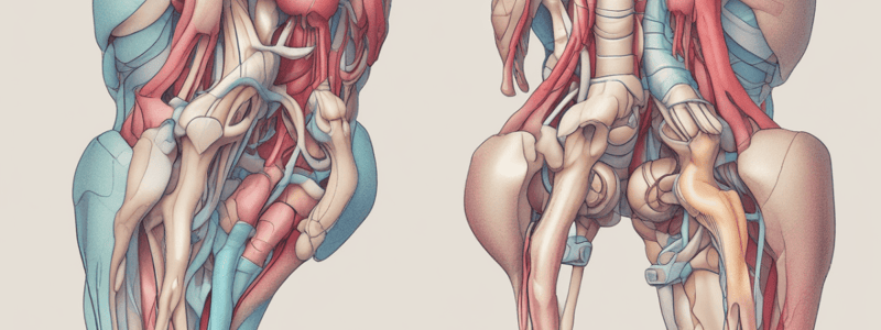
Symphyses
- A symphysis has a pad of fibrocartilage between the articulating bones.
- All symphyses are amphiarthroses, allowing slight mobility.
- Examples of symphyses include:
- The pubic symphysis, located between the right and left pubic bones.
- The intervertebral joints, where the bodies of adjacent vertebrae are separated and united by intervertebral discs.
- Intervertebral discs allow only slight movements between adjacent vertebrae, but collective movements afford the spine considerable flexibility.
Studying That Suits You
Use AI to generate personalized quizzes and flashcards to suit your learning preferences.
Description
Learn about cartilaginous joints, their characteristics, and types, including synchondroses and symphyses. Understand how cartilage connects articulating bones in these joints.


