Podcast
Questions and Answers
Osteoblasts are responsible for the breakdown of bones.
Osteoblasts are responsible for the breakdown of bones.
False
Intramembranous ossification occurs primarily within mesenchymal tissue.
Intramembranous ossification occurs primarily within mesenchymal tissue.
True
Chondrocytes die due to the lack of nutrients when the matrix becomes calcified.
Chondrocytes die due to the lack of nutrients when the matrix becomes calcified.
True
After adolescence, the epiphyseal plate continues to allow for bone growth.
After adolescence, the epiphyseal plate continues to allow for bone growth.
Signup and view all the answers
Flat bones of the skull form through endochondral ossification.
Flat bones of the skull form through endochondral ossification.
Signup and view all the answers
The periosteal cells differentiate into osteoclasts during bone growth in thickness.
The periosteal cells differentiate into osteoclasts during bone growth in thickness.
Signup and view all the answers
During bone growth in length, chondrocytes initially divide and form columns.
During bone growth in length, chondrocytes initially divide and form columns.
Signup and view all the answers
Blood vessels invade the area after osteoclasts dissolve the calcified cartilage.
Blood vessels invade the area after osteoclasts dissolve the calcified cartilage.
Signup and view all the answers
The main function of the skeletal system is to produce hormones for growth and metabolism.
The main function of the skeletal system is to produce hormones for growth and metabolism.
Signup and view all the answers
Hyaline cartilage is the most common type found in joints.
Hyaline cartilage is the most common type found in joints.
Signup and view all the answers
The perichondrium is always present in all types of cartilage.
The perichondrium is always present in all types of cartilage.
Signup and view all the answers
The axial skeleton includes the bones of the skull, vertebral column, and limbs.
The axial skeleton includes the bones of the skull, vertebral column, and limbs.
Signup and view all the answers
Fibrocartilage contains large bundles of collagen and is specifically adapted to resist both compression and tension forces.
Fibrocartilage contains large bundles of collagen and is specifically adapted to resist both compression and tension forces.
Signup and view all the answers
All bones in the human skeleton are classified as long bones.
All bones in the human skeleton are classified as long bones.
Signup and view all the answers
Bone formation can occur through two processes: intramembranous and endochondral ossification.
Bone formation can occur through two processes: intramembranous and endochondral ossification.
Signup and view all the answers
Chondrocytes are the cells found in the lacunae of bone tissue.
Chondrocytes are the cells found in the lacunae of bone tissue.
Signup and view all the answers
Bones primarily serve as a reserve for electrolytes rather than support for body weight.
Bones primarily serve as a reserve for electrolytes rather than support for body weight.
Signup and view all the answers
During the healing of a bone fracture, certain cellular activities are involved, ultimately leading to new bone formation.
During the healing of a bone fracture, certain cellular activities are involved, ultimately leading to new bone formation.
Signup and view all the answers
Bones primarily consist of epithelial tissue.
Bones primarily consist of epithelial tissue.
Signup and view all the answers
The main function of bones is to produce hormones such as osteocalcin.
The main function of bones is to produce hormones such as osteocalcin.
Signup and view all the answers
Long bones have a roughly cuboidal shape.
Long bones have a roughly cuboidal shape.
Signup and view all the answers
Flat bones are thicker than irregular bones.
Flat bones are thicker than irregular bones.
Signup and view all the answers
The vertebrae are classified as irregular bones.
The vertebrae are classified as irregular bones.
Signup and view all the answers
Pneumatized bones contain air pockets.
Pneumatized bones contain air pockets.
Signup and view all the answers
The patella is considered a short bone.
The patella is considered a short bone.
Signup and view all the answers
Sutural bones are flat bones that are irregularly shaped.
Sutural bones are flat bones that are irregularly shaped.
Signup and view all the answers
Blood cell production primarily occurs in the yellow bone marrow.
Blood cell production primarily occurs in the yellow bone marrow.
Signup and view all the answers
Calcium and phosphate can be released into the bloodstream from bones.
Calcium and phosphate can be released into the bloodstream from bones.
Signup and view all the answers
The periosteum is present in sesamoid bones.
The periosteum is present in sesamoid bones.
Signup and view all the answers
The endosteum consists of multiple layers of osteoprogenitor cells.
The endosteum consists of multiple layers of osteoprogenitor cells.
Signup and view all the answers
Compact bone is thicker where mechanical forces are lesser.
Compact bone is thicker where mechanical forces are lesser.
Signup and view all the answers
Bone surface markings for nerve and blood vessel passage are not present at birth.
Bone surface markings for nerve and blood vessel passage are not present at birth.
Signup and view all the answers
The nutrient foramen typically has multiple entry points for blood supply.
The nutrient foramen typically has multiple entry points for blood supply.
Signup and view all the answers
Bones consist of 15% minerals, 30% collagen fibres, and 55% water.
Bones consist of 15% minerals, 30% collagen fibres, and 55% water.
Signup and view all the answers
Osteoblasts are responsible for removing and recycling bone.
Osteoblasts are responsible for removing and recycling bone.
Signup and view all the answers
The internal layer of short bones is primarily composed of spongy bone.
The internal layer of short bones is primarily composed of spongy bone.
Signup and view all the answers
Hydroxyapatite is a mineral that contributes to the rigidity of bone.
Hydroxyapatite is a mineral that contributes to the rigidity of bone.
Signup and view all the answers
Haversian systems are primarily found in the internal layer of spongy bone.
Haversian systems are primarily found in the internal layer of spongy bone.
Signup and view all the answers
The periosteum is composed of an outer fibrous layer and inner cellular layer containing osteoprogenitor cells.
The periosteum is composed of an outer fibrous layer and inner cellular layer containing osteoprogenitor cells.
Signup and view all the answers
Trabeculae in spongy bone are arranged randomly and do not follow lines of stress.
Trabeculae in spongy bone are arranged randomly and do not follow lines of stress.
Signup and view all the answers
Osteocytes are the primary cells responsible for the maintenance and metabolism of healthy bone.
Osteocytes are the primary cells responsible for the maintenance and metabolism of healthy bone.
Signup and view all the answers
Bone marrow is present in short bones but there is no marrow cavity.
Bone marrow is present in short bones but there is no marrow cavity.
Signup and view all the answers
Osteogenic cells are specialized bone cells that do not divide.
Osteogenic cells are specialized bone cells that do not divide.
Signup and view all the answers
Study Notes
Cartilage
- Connective tissue
- Contains collagen and/or elastic fibers in a gel-like structure
- Chondrocytes (cells) are located in lacunae (cavities)
- No nerves or blood vessels
- Surrounded by perichondrium (except articular and fibrocartilage)
- Perichondrium contains fibroblasts (outer layer) and chondroblasts (precursors to chondrocytes, inner layer)
Hyaline Cartilage
- Most common type of cartilage
- Found in most articulations (joints)
Fibrocartilage
- Contains large bundles of collagen
- Resists compression and tension forces
Elastic Cartilage
- Contains a matrix of elastic fibers
- Provides flexibility
Bones
- Organs composed of connective tissues (bone, cartilage, adipose, blood), epithelial tissue, nervous tissue, and blood vessels
- Support body weight
- Allow controlled and precise movements in conjunction with muscles
- Muscles pull against the skeleton for movement.
Bone Functions
- Support: Provide the framework for the body.
- Movement: Act as levers in conjunction with muscles to facilitate movement.
- Protection: Skull, rib cage, vertebral column and pelvis protect vital organs.
- Mineral Storage: Store calcium and phosphate.
- Blood Cell Production and Energy Storage: Red bone marrow produces red and white blood cells.
- Energy Metabolism: Osteocalcin, a hormone produced by bone cells (osteoblasts) influences bone production, fat storage and stimulates insulin production.
Bone Types
- Flat Bones: Thin layers of compact bone surrounding spongy bone (diploë). Examples: roof of the skull, ribs, sternum, scapula.
- Long Bones: Elongated bones. Examples: limbs, fingers, toes.
- Short Bones: Roughly cuboidal shaped bones. Examples: carpals, tarsals.
- Sesamoid Bones: Small, round bones found embedded in tendons. Examples: patella.
- Irregular Bones: Varied shapes. Examples: vertebrae, some facial bones, heel bone.
- Sutural Bones: Oddly shaped bones found between flat bones of the skull.
Composition of Bone Tissue
- Abundant extracellular matrix containing widely separated cells.
- Matrix: 15% water, 30% collagen fibres, 55% minerals
- Collagen fibres provide elasticity.
- Mineral salts provide rigidity (hydroxyapatite, calcium phosphate, magnesium, fluoride, potassium, sulfate).
- Collagen fibres provide an organic framework for mineral crystals to form, resulting in strong, flexible bone.
- Osteogenic (osteoprogenitor) cells: Unspecialized bone cells capable of dividing. Found in the inner and outer linings of bones (endosteum and periosteum).
- Osteoblasts: Bone building cells that secrete collagen fibers and initiate calcification. After surrounding themselves with the matrix, they become osteocytes.
- Osteocytes: Mature bone cells responsible for maintenance and metabolism.
- Osteoclasts: Derived from white blood cells. Located in the endosteum. Involved in bone growth, remodeling and resorption, which regulates calcium levels.
Compact Bone
- External layer (cortex)
- Provides strength and support
- Contains osteons (Haversian systems) which include:
- Lamellae
- Canaliculi
- Central canal
- Osteocytes
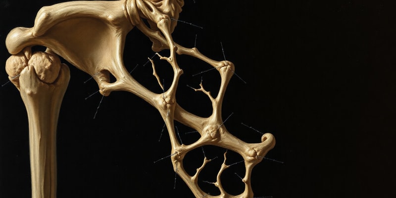
Spongy Bone Tissue
- Internal layer
- Trabeculae have no central canal. Osteocytes in lacunae receive nutrients from blood vessels of the endosteum by diffusion.
- Reduces bone weight
- Supports and protects red bone marrow
- Found in areas with lighter stress
- Trabeculae are arranged along lines of stress
Periosteum
- Outer fibrous layer, inner cellular layer (osteoprogenitor cells).
- Not present in sesamoid bones, at tendon and ligament attachments, joint structures or articular cartilage.
-
Functions:
- Isolates and protects bone from surrounding tissues.
- Provides a route for circulatory and nervous supply.
- Participates in bone growth and repair.
- Anchors the bone to the connective tissue network of the deep fascia.
Endosteum
- Single layer of osteoprogenitor cells
- Active in growth and remodeling
- Covers trabeculae in the medullary cavity or the central canal of osteons
- Not always continuous
Blood and Nerve Supply
- Diaphysis: Nutrient foramen, nutrient artery, nutrient vein.
- Epiphysis and Metaphysis: Several veins and arteries penetrate through foramina.
- Periosteum & outer part: Small veins and arteries branching from the nutrient vein and artery. Perforating canals.
- Nerve supply: Follows veins and arteries. Sensory nerves with many endings in the periosteum and cortical bone, contributing to the pain associated with fractures.
Forces at work
- Compact and spongy bone are aligned along lines of stress.
- Compact bone is thicker where forces are greater, the mesh of spongy bone is also oriented to counteract stress.
- Spongy bone is more appropriate for multidirectional tension.
Bone Surface Markings
- Develop throughout life to offer anchor points for tendons and ligaments as muscles are used (tension and compression forces change the topography).
- Markings that allow the passage of nerves and blood vessels are present from birth.
Studying That Suits You
Use AI to generate personalized quizzes and flashcards to suit your learning preferences.
Related Documents
Description
Test your knowledge about the types of cartilage and their functions, as well as the structure and purpose of bones in the human body. This quiz covers aspects such as hyaline, fibrocartilage, and elastic cartilage, along with bone composition and its vital roles. Challenge yourself on this essential topic in anatomy!