Podcast Beta
Questions and Answers
What is the primary reason osteoporosis is referred to as a 'silent disease'?
Osteoporosis can lead to increased bone density.
False
Name one non-modifiable risk factor for osteoporosis.
Age over 50
Osteoclastic activity is greater than __________ activity in osteoporosis.
Signup and view all the answers
Which demographic is most commonly affected by osteoporosis?
Signup and view all the answers
Match the following types of osteoporosis with their descriptions:
Signup and view all the answers
Exercise can help slow the atrophy of muscle tissue associated with aging.
Signup and view all the answers
What is osteopenia?
Signup and view all the answers
Which of the following individuals is at risk for osteoporosis?
Signup and view all the answers
Osteoarthritis is a chronic metabolic disease.
Signup and view all the answers
Name one medical condition associated with secondary osteoporosis.
Signup and view all the answers
A bone density test may be necessary if you experience height loss of _______ inches or more within one year.
Signup and view all the answers
Match the following conditions with their association with osteoporosis:
Signup and view all the answers
What is one of the clinical manifestations of osteoporosis?
Signup and view all the answers
Both osteoporosis and osteoarthritis can cause height loss.
Signup and view all the answers
What type of exercise is recommended to prevent or lessen the effects of osteoporosis?
Signup and view all the answers
What is the T-score range for osteoporosis?
Signup and view all the answers
A T-score of -2.0 indicates normal bone density.
Signup and view all the answers
Name one pharmacological treatment used for osteoporosis.
Signup and view all the answers
The DEXA scan is also known as _______.
Signup and view all the answers
Match the following types of diagnostic imaging with their descriptions:
Signup and view all the answers
Which of the following is NOT a major risk factor for falls?
Signup and view all the answers
Vitamin D supplementation is a common treatment for osteoporosis.
Signup and view all the answers
What tool is used for fracture risk assessment in osteoporosis patients?
Signup and view all the answers
What is a primary goal of treatment for muscular dystrophy?
Signup and view all the answers
Osteomalacia is characterized by bone softening due to a deficiency in calcium.
Signup and view all the answers
What are two common symptoms of osteomalacia?
Signup and view all the answers
The condition known as __________ occurs in children due to vitamin D deficiency.
Signup and view all the answers
Match the following diagnostic tests with their purpose:
Signup and view all the answers
Which of the following is NOT a recommended treatment for osteomalacia?
Signup and view all the answers
Osteoporosis and osteomalacia are the same condition.
Signup and view all the answers
Name one risk factor for developing osteomalacia.
Signup and view all the answers
What is Dupuytren’s Contracture?
Signup and view all the answers
Hallux Valgus is commonly known as a bunion.
Signup and view all the answers
Name one symptom of Carpal Tunnel Syndrome.
Signup and view all the answers
The primary goal of treatment for Carpal Tunnel Syndrome is to relieve ________ compression.
Signup and view all the answers
Match the types of Muscular Dystrophy with their characteristics:
Signup and view all the answers
What is a common symptom of Muscular Dystrophy?
Signup and view all the answers
Carpal Tunnel Syndrome is primarily caused by hormonal changes.
Signup and view all the answers
What is the primary cause of Plantar Fasciitis?
Signup and view all the answers
Which of the following is NOT a recommended treatment for flexion contractures?
Signup and view all the answers
Mirror therapy can be used for the treatment of muscle shortening.
Signup and view all the answers
What are the signs and symptoms of scoliosis?
Signup and view all the answers
Scoliosis can be diagnosed through a ________, X-ray, ultrasound, or MRI.
Signup and view all the answers
Match the post-operative interventions with their primary focus:
Signup and view all the answers
Which of the following is NOT a classification of osteomyelitis?
Signup and view all the answers
Chronic osteomyelitis typically presents with constant localized pain.
Signup and view all the answers
Name one risk factor for amputation.
Signup and view all the answers
The procedure of __________ involves the surgical removal of a limb.
Signup and view all the answers
Match the following terms with their definitions:
Signup and view all the answers
Which diagnostic tool helps assess arterial circulation for amputation candidates?
Signup and view all the answers
Infection is the only risk factor associated with amputations.
Signup and view all the answers
What is the primary goal of surgical intervention for osteomyelitis?
Signup and view all the answers
In osteomyelitis, __________ formation is a key component of the infection cycle.
Signup and view all the answers
Which of the following is NOT a treatment option for osteomyelitis?
Signup and view all the answers
Which of the following is a modifiable risk factor for osteoporosis?
Signup and view all the answers
Osteoporosis is primarily characterized by increased osteoblastic activity.
Signup and view all the answers
What is the main prevention strategy for osteoporosis?
Signup and view all the answers
Osteoporosis is often referred to as a '________ disease' due to its subtle symptoms.
Signup and view all the answers
Match the following terms related to osteoporosis with their descriptions:
Signup and view all the answers
Which of the following is NOT a symptom typically associated with osteoporosis?
Signup and view all the answers
Caucasian women are the least likely demographic to develop osteoporosis.
Signup and view all the answers
Identify one type of hormone involved in bone metabolism.
Signup and view all the answers
What is the T-score range that indicates osteopenia?
Signup and view all the answers
A DEXA scan is used to measure bone density.
Signup and view all the answers
Name one pharmacological treatment for osteoporosis.
Signup and view all the answers
The major goal of osteoporosis treatment is to reduce the potential for __________.
Signup and view all the answers
Match the following diagnostic tests with their purposes:
Signup and view all the answers
Which of the following factors could increase the risk for osteoporosis in men aged 50-69?
Signup and view all the answers
Which of the following is NOT a major risk factor for falls?
Signup and view all the answers
A height loss of ¾ inch within one year is a potential indicator for needing a bone density test.
Signup and view all the answers
What is the primary purpose of the FRAX tool?
Signup and view all the answers
The treatment for osteoporosis may include __________ therapy to improve bone health.
Signup and view all the answers
Name one medical condition associated with secondary osteoporosis.
Signup and view all the answers
Osteoporosis is primarily characterized by bones becoming less dense and more likely to __________.
Signup and view all the answers
Match the following conditions with their association with osteoporosis:
Signup and view all the answers
Which statement best describes the difference between osteoporosis and osteoarthritis?
Signup and view all the answers
Both osteoporosis and osteoarthritis can lead to pain and height loss.
Signup and view all the answers
List two lifestyle factors that can help prevent osteoporosis.
Signup and view all the answers
Which type of muscular dystrophy typically has an onset in adolescence to early adulthood?
Signup and view all the answers
Duchenne muscular dystrophy affects both males and females equally.
Signup and view all the answers
What is a common symptom of congenital muscular dystrophy?
Signup and view all the answers
Becker muscular dystrophy typically has a survival into __________ age.
Signup and view all the answers
Match each type of muscular dystrophy with its age of onset:
Signup and view all the answers
Which type of muscular dystrophy is characterized by weakness and wasting of shoulder and lower leg muscles with common joint deformities?
Signup and view all the answers
Myotonic muscular dystrophy progresses faster than Limb-Girdle muscular dystrophy.
Signup and view all the answers
What are the symptoms associated with Oculopharyngeal muscular dystrophy?
Signup and view all the answers
In Facioscapulohumeral muscular dystrophy, _______ weakness is one of the key symptoms.
Signup and view all the answers
Match the types of muscular dystrophy to their age of onset:
Signup and view all the answers
Which medical condition is associated with increased interleukins and may affect bone loss?
Signup and view all the answers
Increased levels of thyroid hormone (T4) can lead to decreased osteoclastic activity in bones.
Signup and view all the answers
What condition causes a decrease in estrogen, which is important for bone maintenance?
Signup and view all the answers
Too much __________ increases osteoclastic activity and can contribute to osteoporosis.
Signup and view all the answers
Match the following pathological causes with their associated medical condition:
Signup and view all the answers
Which of the following is a side effect of Denosumab?
Signup and view all the answers
Calcitonin is a synthetic hormone used to increase calcium levels in the body.
Signup and view all the answers
What treatment is contraindicated for women with a history of venous thromboembolism (VTE)?
Signup and view all the answers
The hormone that decreases calcium levels in the body is called _______.
Signup and view all the answers
Which of the following is a sign or symptom associated with Denosumab?
Signup and view all the answers
Match the treatment with its associated function:
Signup and view all the answers
It is advised to get a dental exam before starting Denosumab treatment.
Signup and view all the answers
Which of these side effects is associated with hypercalcemia?
Signup and view all the answers
What should be monitored in patients receiving Denosumab treatment?
Signup and view all the answers
Taking calcium on an empty stomach is recommended for optimal absorption.
Signup and view all the answers
What is the maximum daily dosage of vitamin D for adults over 50 years old?
Signup and view all the answers
Calcium supplementation is recommended to prevent __________ in post-menopausal women.
Signup and view all the answers
Match the following drug names with their primary use:
Signup and view all the answers
Which of the following is NOT a common side effect of bisphosphonates?
Signup and view all the answers
Vitamin D helps in the absorption of calcium.
Signup and view all the answers
What should be monitored before starting calcium supplementation?
Signup and view all the answers
Study Notes
Bone Metabolism Review
- Nutrients - Carbohydrates, Fats, Proteins
- Metabolism - Energy, Cell repair
- Hormones - Chemical Messengers
- Catabolism vs Anabolism
Bone Cells
Bone Remodeling
- Bone remodeling is a continuous process that involves resorption of old bone and formation of new bone
- Resorption is performed by osteoclasts
- Formation is performed by osteoblasts
Aging Bones
- Decreased bone mass and minerals
- Decrease calcium resorption
- Slow resorption of the interior of long bones
- Slow production of new bone on the outside surface of bones
- Vertebrae shorten and intervertebral disks thin, causing kyphosis
- Cartilage on bone surfaces in joints deteriorates and bone spurs may occur
Changes Related to Aging
- Bone structure changes (caused by expansion and resorption)
- Osteopenia - decreased bone density (can lead to osteoporosis)
- Cartilage degeneration (can lead to arthritis)
- Atrophied muscle tissue (increased exercise can slow atrophy)
- Decreased coordination and range of motion
- Loss of muscle strength
- Gait changes
- Slowed movement
- Increased fall risk
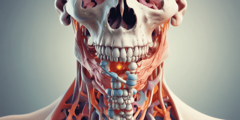
Osteoporosis
- Definition: Chronic metabolic disease in which bone demineralization results in decreased density and subsequent fractures
- Osteoclastic activity is greater than osteoblastic activity
- Most common in postmenopausal, Caucasian women
-
Prevention is key!
- Primary: Prevention measures start early in life with proper nutrition, exercise, and healthy habits
- Secondary: Prevention measures are taken to minimize bone loss after a diagnosis of osteoporosis
- Regional: Prevention measures target specific areas of the body that are at higher risk for osteoporosis, such as the spine or hips
Osteoporosis – Risk Factors
- Non-modifiable: Age greater than 50, Family history, History of low trauma fracture after 50 years old
- Modifiable: Low body weight, thin build, Chronic low calcium or vitamin D intake, Excess or deficient protein intake, Estrogen or testosterone deficiency, Gender confirmation, Smoking, High alcohol intake, Excessive caffeine or carbonated beverage intake
Osteoporosis - Screening
-
Who should have a bone density test?
- Women age 65 or older
- Men age 70 or older
- Anyone who breaks a bone after age 50
- Women of menopausal age with risk factors
- Postmenopausal women under age 65 with risk factors
- Men age 50-69 with risk factors
-
A bone density test may also be necessary if any of the following apply:
- X-ray of the spine shows a break or bone loss in the spine
- Back pain with a possible break in the spine
- Height loss of ½ inch or more in one year
- Total height loss of 1½ inches from original height
Secondary Osteoporosis
- Medical conditions: Diabetes, Hyperthyroidism, Hyperparathyroidism, Cushing's syndrome, Growth hormone deficiency, Metabolic acidosis, Female hypogonadism, Rheumatoid arthritis, Prolonged immobilization, Bone cancer, Cirrhosis
- Medications: Aluminum, Anticonvulsants, Cytotoxic drugs, Glucocorticoids, Gonadotropin-releasing hormone agonists, Immunosuppressants, Lithium, Long-term heparin use, Progesterone, Tamoxifen (postmenopausal use), TPN, Alcohol, Thyroid hormones
Osteoporosis vs. Osteoarthritis
- Osteoporosis: Silent disease, Chronic metabolic disease, Bones become less dense and more likely to fracture, Diet rich in calcium and vitamin D, regular weight-bearing exercise, and healthy lifestyle can prevent or lessen the effects of the disease.
- Osteoarthritis: Painful, Degenerative joint disease, Caused by repeated overuse from performing a particular task or carrying around excess body weight, Treatments aimed at relieving the symptoms of pain and stiffness, Both can cause pain and height loss.
- Many individuals have both.
Osteoporosis – Clinical Manifestations
- Loss of height
- Progressive spinal curvature
- "Dowager's Hump"
- Low back pain
- Fractures
- Constipation
- Reflux
- Respiratory complications
Osteoporosis – Diagnostic Imaging
- X-rays
-
Bone Mineral Density (BMD)
- DEXA Scan: Dual-energy X-ray absorptiometry (T-score on next slide)
- cDXA vs pDXA
- pQUS (peripheral quantitative ultrasound densitometry)
- MRI
- FRAX: Fracture risk assessment tool
DEXA T-Score
- Normal: BMD is within 1 SD of a "young normal" adult (T-score at -1.0 and above)
- Low bone mass ("osteopenia"): BMD is between 1.0 and 2.5 SD below that of a "young normal" adult (T-score between -1.0 and -2.49)
-
Osteoporosis: BMD is 2.5 SD or more below that of a "young normal" adult (T-score at or below -2.5)
- Patients in this group who have already experienced one or more fractures are deemed to have severe or "established" osteoporosis.
Osteoporosis – Diagnostic Labs
- Serum calcium
- Vitamin D
- Alkaline phosphatase
- Phosphorus
- Urinary calcium
- Serum protein
- Thyroid function tests
- Serum & Urinary Bone Turnover Markers
Osteoporosis - Treatment
- Priority problem: Potential for fractures due to weak, porous bone tissue
- Pharmacological treatment: Calcium, Vitamin D, Bisphosphonates, Estrogen Agonists/Antagonists, Monoclonal antibodies/RANKL inhibitors, Calcitonin, Parathyroid hormone-related protein drugs
- Diet therapy: Calcium and vitamin D-rich diet
- Exercise: Weight-bearing exercise and physical therapy
- Pain management: Medications and therapies for pain relief
- Orthotic devices: Braces and supports for stability
- Psychosocial: Counseling and support groups
- Community resources: Home health services and support groups
Osteoporosis - Pharmacology
- Calcium: Essential mineral for bone health, helps to strengthen and maintain bones
- Vitamin D: Promotes calcium absorption from the gut, essential for bone health
-
Bisphosphonates: Inhibit osteoclast activity, reduce bone resorption, decrease fracture risk
- Alendronate (Fosamax): Common bisphosphonate, taken orally
- Risedronate (Actonel): Another common bisphosphonate, taken orally
- Ibandronate (Boniva): Taken orally or intravenously
-
Newer Bisphosphonates:
- IV Zoledronic acid (Reclast): Given intravenously once a year
- IV pamidronate (Aredia): Given intravenously every 3-4 weeks
-
Estrogen Agonists/Antagonists: Inhibit bone resorption, reduce fracture risk
- Raloxifene (Evista): Selective estrogen receptor modulator (SERM), taken orally
-
Monoclonal antibodies/RANKL inhibitors: Target RANK ligand, a protein essential for osteoclast formation and activation, reduce bone resorption
- Denosumab (Prolia): Given as an injection every 6 months
- Calcitonin: Hormone that inhibits osteoclast activity, reduces bone resorption, relieves pain from vertebral fractures
-
Parathyroid hormone-related protein drugs: Stimulate bone formation, increase bone density, reduce fracture risk
- Teriparatide (Forteo): Given as an injection daily
- Abaloparatide (Tymlos): Given as an injection once a day
Caregiver Teaching - Fall Prevention
- Major risk factors: Delirium, dementia, immobility, muscular weakness, history of falls, visual or hearing deficits, current medications, environmental hazards, malnutrition, agitation
- Remove rugs: Reduce tripping hazards
- Place adequate stair rails and grab bars: Provide support and safety
- Improve shower/bath access: Eliminate potential slipping hazards
- Use of orthotic devices: Assistive devices to improve balance and stability
Hand & Foot Disorders
- Dupuytren's Contracture: Thickening and tightening of tissue under the skin of the hand, causing a gradual bending of the fingers.
- Ganglion Cyst: Small, round, fluid-filled sac growing out of the tissues around a joint.
- Hallux Valgus (Bunion): The big toe drifts laterally and the first metatarsal head becomes enlarged.
- Hammer Toe: The toe is bent at the middle joint, causing a hammer-like appearance.
- Plantar Fasciitis: Inflammation of the plantar fascia, a thick band of tissue on the bottom of the foot.
-
Carpal Tunnel Syndrome (CTS): Compression of the median nerve in the wrist, causing numbness, tingling, and weakness in the hand.
- Common causes: Repetitive stress, genetics, and rheumatoid arthritis.
- Assessment: Pain, numbness, tingling, motor changes.
- Diagnosis: Physical exam, Phalen maneuver, Tinel's sign, X-ray, MRI, Ultrasound, and nerve conduction studies.
-
Treatment: Relieve pressure on the median nerve.
- Medication: NSAIDs, steroids, diuretics.
- Immobilization: Wrist splints.
- Surgery: Endoscopic carpal tunnel release (ECTR), open carpal tunnel release (OCTR), synovectomy.
- Post-op care: Monitor vital signs, dressing, pain relief, NV checks, wrist splint, lifting restrictions, home care.
Muscular Dystrophies
-
Muscular Dystrophy (MD): A group of genetic diseases that cause progressive muscle weakness and loss of muscle mass.
- Types: Duchenne, Becker, Limb-Girdle, Facioscapulohumeral, Myotonic, Congenital, Distal, Emery-Dreifuss, Oculopharyngeal.
-
Duchenne & Becker MD: X-linked disorders that differ in severity.
- Cause: Mutation in the dystrophin gene.
- Manifestations: Muscle weakness starting early in life, frequent falls, waddling gait, lordosis, Gower's sign (using hands to stand up), pseudohypertrophy (enlargement of muscles due to fat and connective tissue), cardiomyopathy, cognitive impairment.
- Duchenne: Most severe, onset 2-6 years old, loss of mobility by age 12, rare to survive past 20 years old.
- Becker: Less severe than Duchenne, onset in adolescence or early adulthood.
- Diagnosis: Physical exam, genetic testing, muscle biopsy, neurological tests, serum creatine kinase, myoglobin.
-
Treatment: No cure, but many clinical trials ongoing.
- Primary goal: Maintain optimal functioning.
- Secondary goal: Prevent contractures.
- Treatment methods: Physical therapy, corticosteroids, stretching, strength and muscle training, breathing exercises, ROM exercises, surgery, respiratory and cardiac support (noninvasive ventilation, tracheostomy, mechanical ventilation, extensive cardiac evaluation), palliative care.
Osteomalacia
-
Osteomalacia: Softening of the bones due to deficiency or malabsorption of vitamin D.
- Also known as Rickets in children.
- Risk Factors: Decreased sunlight exposure, vitamin D deficiency, malabsorption syndromes, dark skin, obesity, elderly, medications, renal or hepatic disease.
- Clinical Manifestations: Hypocalcemia, pain (especially in arms, legs, and spine), pain worse at night, waddling gait, decreased muscle tone, fractures, bowlegs, pigeon chest, restlessness, fever.
- Diagnosis: X-ray, bone mineral density scan, blood and urine tests.
- Treatment: Supplements (vitamin D, calcium, phosphorus), increase sunlight exposure, dietary changes, corrective braces, surgery to correct bone deformities, routine lab draws.
Osteomyelitis
- Severe bone and surrounding tissue infection requiring immediate treatment.
- Infection Cycle: Pathogen invasion leads to tissue inflammation, edema formation, decreased blood flow, bone necrosis, and abscess formation.
-
Classification:
- Exogenous: Infection from outside the body.
- Endogenous: Infection carried by the bloodstream.
- Contiguous: Infection spreads from adjacent tissue.
-
Acute vs. Chronic:
- Acute: High fever, swelling, erythema, tenderness, constant localized pain, intensified pain during movement.
- Chronic: Skin ulceration, sinus tract formation, constant localized pain, drainage.
-
Assessment:
- Elevated vital signs, including expected fever.
- Pain, tenderness, and inflammation.
- Signs and symptoms of infection.
- Vomiting and dehydration.
- Limping or hesitation.
- Malaise.
- Drainage.
-
Diagnostics:
- Labs: Blood cultures and wound cultures.
- Imaging: X-ray, needle aspiration, open bone biopsy, radionuclide bone scans, MRI.
-
Treatment:
- Antibiotic therapy: Home-based or outpatient.
- Contact Precautions/Infection Control: Implement infection control measures.
- Wound care: Manage wound, promoting healing.
- Pain control: Address pain effectively.
- Hyperbaric O2 Therapy: Potential treatment for osteomyelitis.
-
Surgical intervention:
- Sequestrectomy: Removal of infected bone fragments.
- PMMA beads: Bone cement for stabilization.
- Bone grafts: Replacement of infected bone with healthy bone.
- Muscle Flaps: Surgical technique to cover and protect exposed bone.
- Amputation: Extreme measure when other therapies fail.
Amputation
-
Types:
- Congenital: Present at birth.
- Surgical: Performed due to medical reasons.
- Traumatic: Resulting from injury.
-
Risk Factors:
- Diabetes mellitus.
- Peripheral Vascular Disease (PVD).
- Arteriosclerosis.
- Infection.
- Cancer.
- Joint dysfunction.
-
Traumatic Amputations - Field Care:
- ABCs: Assess airway, breathing, and circulation.
- Salvage the body part: If possible, preserve the amputated limb.
- Promote Perfusion: Maintain blood flow to the injured area.
- Prevent hemorrhage: Control bleeding effectively.
- Transport to hospital ASAP: Ensure timely medical care.
-
(Pre)Amputation Assessment:
- Health History: Gather comprehensive patient information.
- Subjective: Patient's description of symptoms and concerns.
- Objective: Observable physical findings and measurements.
- Psychosocial: Assess emotional and social well-being.
- Knowledge Deficits: Identify areas where patient requires education.
-
(Pre)Amputation Diagnostics:
- Ankle-brachial index (ABI): Measures blood flow in the lower limbs.
- Doppler ultrasound: Uses sound waves to visualize blood flow.
- Laser doppler flowmetry: Non-invasive method to assess blood flow.
-
ABI Value Interpretation:
- Greater than 1.4: Possible calcification or vessel hardening. Referral to vascular specialist recommended.
- 1.0-1.4: Normal. No action required.
- 0.9-1.0: Acceptable. No action required.
- 0.8-0.9: Some arterial disease. Treat risk factors.
- 0.5-0.8: Moderate arterial disease. Referral to vascular specialist recommended.
- Less than 0.5: Severe arterial disease. Referral to vascular specialist recommended.
-
Amputation Surgery:
- Aims for successful healing, functional preservation, and prosthetic ability.
- Standard Amputation: Traditional surgical technique.
- Osseointegration: Implantation of a prosthesis directly into bone.
- Interdisciplinary: Collaboration among medical professionals.
-
Amputation - Lower Extremity:
- Partial foot amputation: Toe, midfoot, or Syme amputation.
- Syme Ankle disarticulation: Removal of the foot at the ankle joint.
- Below-the-knee amputation: Removal of the foot and part of the lower leg.
- Knee disarticulation: Removal of the lower leg at the knee joint.
- Above-the-knee amputation: Removal of the lower leg and part of the upper leg.
- Hip disarticulation: Removal of the entire leg at the hip joint.
-
Amputation - Upper Extremity:
- Partial hand amputation: Removal of part of the hand.
- Wrist disarticulation: Removal of the hand at the wrist joint.
- Below-the-elbow amputation: Removal of part of the lower arm.
- Elbow disarticulation: Removal of the lower arm at the elbow joint.
- Above-the-elbow amputation: Removal of the lower arm and part of the upper arm.
- Shoulder disarticulation: Removal of the entire arm at the shoulder joint.
- Forequarter amputation: Removal of the arm and part of the shoulder girdle.
-
Amputation - Surgery Complications:
- Hemorrhage: Excessive bleeding.
- Infection: Microbial contamination of the surgical wound.
- Impaired mobility: Reduced range of motion and difficulty moving.
- Neuromas: Benign nerve tumors that cause pain.
- Flexion contractures: Shortening of muscles or tendons, hindering limb function.
- Phantom limb pain: Pain perceived in the missing limb.
-
Phantom Limb Pain:
- Common occurrence following amputation.
- Real pain with various triggers.
- Interferes with activities of daily living (ADLs).
-
Treatment:
- Physical Therapy (PT), Massage, Heat, Transcutaneous Electrical Nerve Stimulation (TENS): Non-pharmacological approaches.
- Mirror therapy: Visual illusion used to reduce pain.
-
Pharmacological:
- IV Calcitonin: Calcitonin hormone for pain relief.
- Beta-blockers: Medications to reduce heart rate and blood pressure.
- Anticonvulsants: Medications used to control seizures.
- Antispasmodics: Medications to relieve muscle spasms.
- Opioids: Pain relievers.
- Antidepressants: Medications used to treat depression.
- NMDA antagonists: Medications that block the action of N-methyl-D-aspartate (NMDA) receptors.
-
Flexion Contractures:
- Muscle or tendon shortening.
- Potential obstacle for prosthetic limb use.
-
Prevention:
- Exercises: Stretching and strengthening exercises.
- Splinting: Devices to immobilize and support the limb.
- Avoid pillow under the knee: Minimize knee flexion.
-
Post-op Interventions:
- Assess and maintain tissue perfusion: Monitor blood flow to the surgical area.
- Pain management: Provide effective pain relief.
- Prevent infection, promote wound healing: Implement infection control measures and support healing.
- Improve mobility: Encourage and facilitate physical activity.
- Prostheses preparation: Prepare for prosthetic limb fitting and use.
- Promotion of body image: Address the psychological impact of amputation.
- Lifestyle adaptations: Assist with adapting to daily life after amputation.
-
Amputation - Prosthetics:
- Preoperative preparation: Prepare for prosthetic fitting.
- Postoperative considerations: Prevent secondary disabilities, ensure proper prosthetic fit, manage stump conditioning and shrinking, facilitate rehabilitation, and provide education.
Scoliosis
-
Assessment:
- Signs and Symptoms: Uneven shoulders, prominent shoulder blade, uneven waist, uneven hips, one side of the rib cage jutting forward, prominence on one side of the back when bending forward.
-
Diagnosis:
- Physical Exam: Adams forward bend test, plumb line test, scoliometer.
- Imaging: X-ray, ultrasound, MRI to confirm diagnosis.
-
Treatment:
- Close monitoring with x-rays: Regular imaging for progress monitoring.
- Brace: Used to correct and support the spine.
-
Surgery: Employed in more severe cases.
- Spinal fusion: Joining vertebrae to stabilize the spine.
- Expanding rod: Device for gradual spinal correction.
- Vertebral body tethering: Procedure to tether the spine and promote growth.
Bone Metabolism
- Bone metabolism is the process of continuous growth and remodeling
- Bone growth is a complex and regulated process involving cells such as osteoblasts, which build bone, and osteoclasts, which break down bone
- Bone remodeling is constantly working to maintain bone health and strength.
Bone Cells
- Osteoblasts are the cells responsible for building new bone tissue. Responsible for bone formation.
- Osteoclasts are responsible for the breakdown and resorption of bone tissue. Responsible for bone resorption.
Aging Bones
- Aging bones experience a decrease in bone mass and minerals.
- Calcium resorption slows, leading to a slower rate of bone formation.
- Vertebrae shorten and intervertebral discs thin. Kyphosis can develop.
- Cartilage on bone surfaces within joints deteriorate and bone spurs may occur.
Changes Related to Aging
- Bone structure changes, expansion, and resorption.
- This leads to osteopenia, a condition where the density of bone is decreased.
- Cartilage degeneration can lead to arthritis.
- Atrophy of muscle tissue can occur but can be slowed with increased exercise.
- Decreased coordination and range of motion is common.
- Loss of muscle strength can increase fall risk.
- Gait changes and slowed movement.
- Increased fall risk.
Osteoporosis
- Chronic metabolic disease where bone demineralization decreases bone density and increases fracture risk.
- Osteoporosis is often referred to as a "silent disease" because it may not present with symptoms until a fracture occurs.
- Osteoclasts dominate osteoblasts, resulting in more bone breakdown than formation.
- More common in postmenopausal women, particularly Caucasian women.
- Prevention is of the utmost importance!
- Three types: primary, secondary, and regional.
Osteoporosis - Risk Factors
- Non-modifiable*
- Age: greater than 50 years old.
- Family history of osteoporosis.
- History of low-trauma fractures after 50 years old.
- Modifiable*
- Low body weight and thin build.
- Chronic low calcium or vitamin D intake.
- Excess or deficient protein intake.
- Estrogen or testosterone deficiency.
- Gender confirmation.
- Smoking.
- High alcohol intake.
- Excessive caffeine or carbonated beverage intake.
Osteoporosis - Screening
- Women age 65 or older should have a bone density test.
- Men age 70 or older should have a bone density test.
- Anyone who breaks a bone after age 50 should have a bone density test.
- Women of menopausal age with risk factors should have a bone density test.
- Postmenopausal women under age 65 with risk factors should have a bone density test.
- Men aged 50-69 with risk factors should have a bone density test.
- Individuals with an X-ray of the spine showing a break or bone loss in the spine should have a bone density test.
- Individuals with back pain that might indicate a fracture of the spine should have a bone density test.
- Individuals who have experienced a loss of height of ½ inch or more within a year or a total height loss of 1½ inches from their original height should have a bone density test.
Secondary Osteoporosis
-
Medical conditions associated with secondary osteoporosis include:
- Diabetes.
- Hyperthyroidism.
- Hyperparathyroidism.
- Cushing's syndrome.
- Growth hormone deficiency.
- Metabolic acidosis.
- Female hypogonadism.
- Rheumatoid arthritis.
- Prolonged immobilization.
- Bone cancer.
- Cirrhosis.
-
Medications associated with secondary osteoporosis include:
- Aluminum.
- Anticonvulsants.
- Cytotoxic drugs.
- Glucocorticoids.
- Gonadotropin-releasing hormone agonists.
- Immunosuppressants.
- Lithium.
- Long-term heparin use.
- Progesterone.
- Tamoxifen (postmenopausal use).
- TPN.
- Alcohol.
- Thyroid hormones.
Osteoporosis vs Osteoarthritis
-
Osteoporosis: Silent disease that causes bones to become less dense and more brittle. It is a chronic metabolic disease. Diet rich in calcium and vitamin D, regular weight-bearing exercise, and a healthy lifestyle can prevent or lessen the effects of the disease.
-
Osteoarthritis: Painful degenerative joint disease that causes cartilage to wear away. It is caused by repeated overuse of a joint. Treatments aim to relieve the pain and stiffness caused by the disease.
-
Osteoporosis and osteoarthritis can both cause pain and height loss. Some patients have both conditions.
Osteoporosis - Clinical Manifestations
- Loss of height.
- Progressive spinal curvature, which can lead to a "Dowager's Hump."
- Low back pain.
- Fractures.
- Constipation.
- Reflux.
- Respiratory complications.
Osteoporosis - Diagnostic Imaging
- Imaging*
- X-rays: Can reveal fractures and bone density changes.
-
Bone Mineral Density (BMD): Measures bone density using specialized X-ray equipment.
-
DEXA Scan: Dual-energy X-ray absorptiometry is the gold standard for measuring bone density. T-scores are used to assess bone health.
- cDXA: Central DEXA scan which evaluates the lumbar spine and hip.
- pDXA: Peripheral DEXA scan that measures bone density in the forearm or heel.
- pQUS (peripheral quantitative ultrasound densitometry): A test to measure bone density in the heel using ultrasound.
-
DEXA Scan: Dual-energy X-ray absorptiometry is the gold standard for measuring bone density. T-scores are used to assess bone health.
- MRI: Can be used to visualize soft tissues and assess cartilage health.
- FRAX*: Fracture Risk Assessment Tool used to calculate an individual's 10-year risk of major osteoporotic fractures.
Osteoporosis - Diagnostic Labs
- Serum calcium: Measures the amount of calcium in the blood.
- Vitamin D: Measures the amount of vitamin D in the blood.
- Alkaline phosphatase: Enzyme primarily found in bone, liver, and intestines. An elevation may indicate bone formation.
- Phosphorus: Measures the amount of phosphorus in the blood.
- Urinary calcium: Measures the amount of calcium excreted in the urine.
- Serum protein: Measures the amount of protein in the blood.
- Thyroid function tests: Assess thyroid hormone levels to rule out hyperthyroidism as a factor.
- Serum & Urinary Bone Turnover Markers: Measure the rate of bone formation and breakdown, providing information about the activity of osteoblasts and osteoclasts.
Osteoporosis - Treatment
-
Priority problem:* Potential for fractures due to weak, porous bone tissue.
-
Pharmacological treatment:
- Calcium: Supplementation is often recommended to increase calcium intake.
- Vitamin D: Supplementation is often recommended to enhance calcium absorption.
-
Bisphosphonates: These medications slow bone breakdown and increase bone density.
- Alendronate (Fosamax): Oral medication.
- Risedronate (Actonel): Oral medication.
- Ibandronate (Boniva): Oral medication.
-
Newer Bisphosphonates
- IV Zoledronic acid (Reclast): Given by IV once a year.
- IV pamidronate (Aredia): Given by IV every 3-4 weeks.
-
Estrogen Agonists/Antagonists:
- Raloxifene (Evista): Oral medication that can help prevent bone loss.
-
Monoclonal Antibodies/RANKL Inhibitors
- Denosumab (Prolia): Given by injection every six months that prevents bone breakdown by targeting RANKL, a molecule involved in bone breakdown.
-
Calcitonin: An injectable or nasal spray medication that helps slow bone breakdown.
-
Parathyroid hormone-related protein drugs:
- Teriparatide (Forteo): An injection given daily that stimulates new bone growth.
- Abaloparatide (Tymlos): A daily injection that stimulates new bone growth.
-
Diet therapy: A diet rich in calcium and vitamin D is recommended.
-
Exercise: Weight-bearing exercise and resistance training is essential to promote bone health.
-
Pain Management: Medications for pain relief.
-
Orthotic devices: Braces or splints used to support bones and prevent fractures.
-
Psychosocial: Provide emotional support and education.
-
Community Resources: Connect individuals with community resources for support and assistance.
Caregiver Teaching on Fall Prevention
-
Major Risk Factors:
- Delirium.
- Dementia.
- Immobility.
- Muscular weakness.
- History of falls.
- Visual or hearing deficits.
- Current medications.
- Environmental.
- Malnutrition.
- Agitation.
-
Preventative measures:
- Remove throw rugs and clutter.
- Ensure adequate stair rails and grab bars.
- Assess shower and bath access for safety.
- Use of orthotic devices as needed.
Muscular Dystrophy Types and Characteristics
-
Becker Muscular Dystrophy
- Onset: Adolescence to early adulthood
- Symptoms: Similar to Duchenne, but less severe; slower progression
- Life Expectancy: Survival into middle age, almost always affects males
-
Congenital Muscular Dystrophy
- Onset: Birth
- Symptoms: General muscle weakness, potential joint deformities
- Progression: Slow
- Life Expectancy: Shortened lifespan
-
Duchenne Muscular Dystrophy
- Onset: 2 to 6 years
- Symptoms: General muscle weakness and wasting, affecting pelvis, arms, and legs; eventually all voluntary muscles
- Progression: Rapid, often leading to death before the age of 20
- Life Expectancy: Beyond 20's is rare, primarily affects males, but women can be affected with milder symptoms and better prognosis
-
Distal Muscular Dystrophy
- Onset: 40 to 60 years
- Symptoms: Weakness and wasting in hands, forearms, and lower legs
- Progression: Slow
- Rarely leads to total incapacity
-
Emery-Dreifuss Muscular Dystrophy
- Onset: Childhood to early teens
- Symptoms: Weakness and wasting in shoulders, upper arms, and shins; joint deformities
- Progression: Slow; potential for sudden death due to cardiac problems
-
Facioscapulohumeral Muscular Dystrophy
- Onset: Childhood to early adulthood
- Symptoms: Facial muscle weakness, weakness and wasting in shoulders and upper arms
- Progression: Slow with periods of rapid deterioration
- Life Expectancy: Many decades after onset
-
**Limb-Girdle Muscular Dystrophy **
- Onset: Late childhood to middle age
- Symptoms: Weakness and wasting, affecting shoulder and pelvic girdle first
- Progression: Slow
- Cause of Death: Usually cardiopulmonary complications
-
Myotonic Muscular Dystrophy
- Onset: 20 to 40 years
- Symptoms: Weakness in all muscle groups, delayed muscle relaxation after contraction
- Progression: Slow, can span 50 to 60 years
- Areas Affected: Face, feet, hands, and neck
-
**Oculopharyngeal Muscular Dystrophy **
- Onset: 40 to 70 years
- Symptoms: Eyelid and throat muscle weakness leading to swallowing difficulty and emaciation
- Progression: Slow, but eventually causes swallowing difficulties and emaciation as a result of food deprivation
Lack of Activity
- Decreased bone formation due to lack of physical activity
- Vitamin D deficiency due to insufficient sunlight exposure
### Hyperthyroidism
- Increased osteoclastic activity (bone breakdown) due to excessive thyroid hormone (T4)
### Hypothyroidism
- Less osteoblastic activity due to a deficiency in thyroid hormone
### Diabetes
- Decreased osteoblastic activity due to poor glycemic control
### Cushing's Syndrome
- Increased osteoclastic activity due to excessive cortisol
### Hyperparathyroidism
- Increased osteoclastic activity (bone breakdown) due to excessive parathyroid hormone (PTH)
### Hypoparathyroidism
- Decreased vitamin D absorption
- Imbalance of bone turnover caused by a lack of parathyroid hormone (PTH)
### Osteogenesis Imperfecta
- Bone breakdown from the disease process
- Bone breakdown from chemotherapy agents
### Rheumatoid Arthritis
- Inflammation impairs calcium and vitamin D absorption
- Corticosteroids medication promotes osteoclastic activity
### Crohn's Disease
- Inflammation impairs calcium and vitamin D absorption
- Increased Interleukins and tumor necrosis factor - may lead to accelerated bone loss
- Protease inhibitors and antiretroviral therapy (ART) therapies negatively impact osteoblasts
### Multiple Myeloma
- Increased Interleukins and tumor necrosis factor - may lead to accelerated bone loss
- Protease inhibitors and antiretroviral therapy (ART) therapies negatively impact osteoblasts
### Menopause
- Decreased estrogen levels contribute to osteoporosis
- Estrogen is essential for bone density maintenance
Selective Estrogen Receptor Modulators (SERMs)
- Raloxifene is a SERM that mimics estrogen in some parts of the body while blocking its effects elsewhere
- Contraindicated in women with a history of venous thromboembolism (VTE), women who are not menopausal
- Common side effects may include trouble sleeping, spotting, and increased risk of VTE, especially in the first four months of treatment
Monoclonal Antibodies/RANK Ligand Inhibitors
- Denosumab, a monoclonal antibody, is a RANK ligand inhibitor that blocks proteins to decrease bone loss, increase bone mass, and prevent osteoporosis
- Denosumab is commonly used when other treatments have failed to show improvement
- Potential side effects include low Calcium levels, back pain, increased cholesterol, increased risk of urinary tract infections, and maxillary necrosis
- Patients should monitor calcium, magnesium, and phosphorus levels
- Obtain a dental examination before starting treatment
- Important to report new muscle, skeletal pain, and signs of infection
Calcitonin
- Calcitonin is a synthetic parathyroid hormone that decreases calcium levels and increases bone density.
- It is commonly used in postmenopausal women after five years of menopause
- Available as a sublingual spray, subcutaneous injection, or synthetic pill
- Common side effects include epistaxis (nosebleeds), and rhinitis
- Patients should monitor calcium levels for hypocalcemia, and be aware of fish allergies (salmon)
Parathyroid hormone (PTH) related Drugs
- PTH related drugs are injectable and used for patients taking other medication.
Calcium
- Recommended calcium intake is 1000 mg/day for ages 50-70, and 1200 mg/day for ages 71 and older
- Maximum recommended intake is 2500 mg/day
- Common side effects include constipation, cramps, nausea, vomiting, metallic taste, kidney stones, hypercalcemia
- To reduce side effects, intake should be split throughout the day and taken with 6-8 glasses of water
Vitamin D
- Recommended Vitamin D intake is 800-1000 IU for individuals over age 50, with a maximum intake of 4000 IU per day
- Vitamin D helps absorb calcium
- May also be used for babies who are exclusively breastfeeding to help prevent osteomalacia (rickets)
- Overdose may cause hypercalcemia, resulting in bone pain, lethargy, anorexia, nausea, vomiting, increased urination, hallucinations, and dysrhythmias
Bisphosphonates
- Bisphosphonates are natural substances that inhibit bone breakdown used for the prevention and treatment of osteoporosis
- Commonly used bisphosphonates include alendronate, ibandronate, risedronate, pamidronate (IV), and zoledronic acid (IV)
- Individuals should take bisphosphonates for two years, undergo a bone mineral density (DXA) scan, and continue taking for up to five years if bone loss persists
- Common side effects include nausea, vomiting, upset stomach, diarrhea, osteonecrosis, bone pain, back pain, bone fractures, gastric ulcers, esophageal perforations, anemia, osteonecrosis of the jaw, and atrial fibrillation
- Contraindicated in patients with poor renal function, hypocalcemia, and gastroesophageal reflux disease (GERD)
- Take on an empty stomach first thing in the morning with a full glass of water
- Sit upright for 30 minutes after taking the medication to prevent esophageal inflammation.
- Do not give to patients sensitive to aspirin
- The effectiveness of bisphosphonates is increased within the first year of treatment, but plateaus after 2-3 years
- Monitor calcium and kidney function levels
- Obtain a dental exam before starting treatment
Estrogen Agonists/Antagonists
- Used to treat osteoporosis in postmenopausal women
- Potential side effects include blood clots, DVT, hot flashes
- Monitor liver function and obtain AST/ALT levels.
Studying That Suits You
Use AI to generate personalized quizzes and flashcards to suit your learning preferences.
Related Documents
Description
Explore the intricate processes of bone metabolism, including the roles of nutrients, hormones, and bone cells. Learn about bone remodeling, aging effects on bone mass, and the changes that can lead to conditions like osteopenia and osteoporosis. This quiz will deepen your understanding of the physiological aspects of bone health.