Podcast Beta
Questions and Answers
Which ligament is specifically responsible for preventing hyperextension of the hip joint?
What type of joint is the hip joint classified as?
Which structure provides a protective and supportive function to the hip joint?
Which ligament prevents excessive abduction and extension at the hip joint?
Signup and view all the answers
Which articulation occurs at the hip joint?
Signup and view all the answers
What is the primary function of the great saphenous vein?
Signup and view all the answers
Which lymph nodes are likely to become painful due to an inflammatory process in the lower extremities?
Signup and view all the answers
What occurs when there is inflammation in the lower extremities?
Signup and view all the answers
Where does the short saphenous vein primarily drain blood from?
Signup and view all the answers
What is the role of deep fascia in the lower extremities?
Signup and view all the answers
What anatomical structure does the cribriform fascia primarily relate to?
Signup and view all the answers
Which cutaneous nerve is responsible for supplying the skin of the lower lateral quadrant of the buttocks?
Signup and view all the answers
What lies in front of the knee and comprises the terminal branches of cutaneous nerves?
Signup and view all the answers
Which nerve is a branch of the femoral nerve that descends down to innervate the patellar area?
Signup and view all the answers
Which of the following is NOT mentioned as a terminal branch of the lumbar plexus?
Signup and view all the answers
Which of these structures is involved in the patellar plexus?
Signup and view all the answers
Which cutaneous nerve supplies the medial part of the thigh?
Signup and view all the answers
What is the primary role of the infrapatellar branch of the saphenous nerve?
Signup and view all the answers
What is the primary action performed by the adductor longus muscle?
Signup and view all the answers
Which nerve is responsible for innervating both the adductor longus and adductor brevis muscles?
Signup and view all the answers
Where does the adductor brevis originate?
Signup and view all the answers
What is associated with the anterior division of the obturator nerve?
Signup and view all the answers
Which structure is considered an intracapsular ligament of the hip joint?
Signup and view all the answers
What is the main action of the muscles innervated by the posterior division of the obturator nerve?
Signup and view all the answers
What is the insertion point of the adductor longus muscle?
Signup and view all the answers
What common action do the sartorius and adductor longus muscles share?
Signup and view all the answers
What structure is formed by the bony margins of the acetabular notch and completed by the transverse ligament of the hip?
Signup and view all the answers
Which muscle assists in the adduction of the hip joint along with the adductor magnus?
Signup and view all the answers
What movement is limited by the tension in the ligamentum teres femoris?
Signup and view all the answers
Which muscle is NOT involved in lateral rotation of the hip?
Signup and view all the answers
What does the capsule of the hip joint primarily protect?
Signup and view all the answers
Which ligament passes through the space behind the transverse acetabular ligament?
Signup and view all the answers
Which of the following describes the attachment points of the capsule of the hip joint?
Signup and view all the answers
Which muscle is primarily responsible for the lateral rotation of the hip joint?
Signup and view all the answers
Study Notes
ANTEROMEDIAL THIGH & HIP JOINT
-
Cribriform fascia: a deep layer of superficial fascia, it is located over the fossa ovalis and iliac crest
-
Cutaneous Nerve:
- Patellar plexus: located in front of the knee. Includes terminal branches of the lateral, intermediate, and medial cutaneous nerves of the thigh, a terminal branch of the lumbar plexus and femoral nerve, and the infrapatellar branch of the saphenous nerve.
- Saphenous Nerve: another branch of the femoral nerve that descends down, innervating the patellar area.
-
Superficial Lymph nodes:
- Divided into horizontal and vertical groups
- Located along the inguinal ligament and are responsible for draining the lower extremities
-
Deep Fascia:
- Covers the muscles
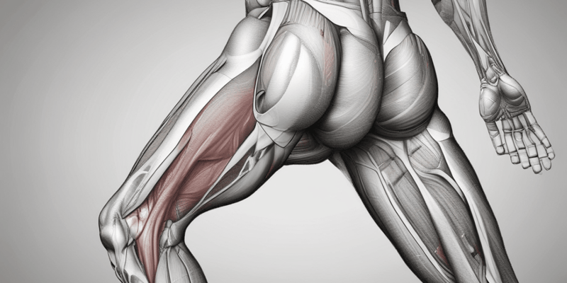
OBTURATOR NERVE & VESSELS
-
Obturator Nerve Arises from the lumbar plexus (anterior division of L2, L3, L4 spinal nerves) and emerges at the medial border of the psoas muscle within the abdomen.
- Leaves the pelvis through the obturator foramen and divides into anterior and posterior divisions
- Anterior Division: Innervates adductor longus, brevis, and gracilis.
- Posterior Division: Supplies adductor magnus.
- Leaves the pelvis through the obturator foramen and divides into anterior and posterior divisions
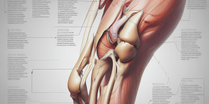
HIP JOINT
- Type: Synovial, Ball & Socket joint
- Diarthrosis: Freely movable joint
- Articulating bones: Head of femur & acetabulum
-
Capsule: Attached to the:
- Acetabular labrum medially
- Intertrochanteric line & neck of femur laterally
- Protective & supportive parts: Joint capsule, synovial membrane, synovial fluid & cavity, ligaments
HIP JOINT LIGAMENTS
-
Intracapsular Ligaments: Found inside the joint capsule
- Ligament of head of femur: Only intracapsular ligament, a small structure that runs from the acetabulum to the fovea capitis of the femur.
-
Extracapsular Ligaments: Found outside the capsule
- Iliofemoral ligament/ Y-ligament of Bigelow: 2 strips of ligaments, coming from the ilium and connected to the base of the neck of femur. It prevents hyperextension of the hip joint.
- Pubofemoral ligament: Coming from the pubis and connected to the femur. It prevents abduction and extension.
- Ischiofemoral ligament: Coming from the ischium and connected to the base of the neck of femur. It prevents hyper extension.
HIP JOINT CAPSULE
- Protects the ball & socket joint
- Attached to the:
- Acetabular labrum above
- Base of neck of femur distally
Studying That Suits You
Use AI to generate personalized quizzes and flashcards to suit your learning preferences.
Related Documents
Description
Test your knowledge of the anatomy of the anteromedial thigh and hip joint, focusing on key structures such as the cribriform fascia, cutaneous nerves, and obturator nerve. This quiz covers important lymph nodes, nerves, and their functions in the lower extremity. Perfect for students studying human anatomy and physiology.