Podcast
Questions and Answers
Which nerve innervates the medial thigh?
Which nerve innervates the medial thigh?
- Femoral nerve (L2-L4)
- Sciatic nerve (L4-S3)
- Obturator nerve (L2-L4) (correct)
- Tibial nerve (L4-S3)
Which nerve is responsible for motor innervation of the posterior thigh and leg, as well as the plantar foot?
Which nerve is responsible for motor innervation of the posterior thigh and leg, as well as the plantar foot?
- Tibial nerve (L4-S3) (correct)
- Common peroneal nerve (L4-S2)
- Obturator nerve (L2-L4)
- Femoral nerve (L2-L4)
Which nerve supplies the perineum and travels with the internal pudendal artery?
Which nerve supplies the perineum and travels with the internal pudendal artery?
- Pudendal nerve (S2-S4) (correct)
- Sciatic nerve (L4-S3)
- Obturator nerve (L2-L4)
- Posterior femoral cutaneous nerve (S1-S3)
Which sacral plexus nerve provides sensory innervation to the posterior thigh?
Which sacral plexus nerve provides sensory innervation to the posterior thigh?
Which nerve is responsible for motor innervation of the anterior and lateral leg, as well as the dorsal foot?
Which nerve is responsible for motor innervation of the anterior and lateral leg, as well as the dorsal foot?
Which division of the lumbar plexus innervates the anterior thigh?
Which division of the lumbar plexus innervates the anterior thigh?
According to Hilton's Law, which of the following is true?
According to Hilton's Law, which of the following is true?
Which of the following nerves is responsible for the sensory innervation of the hip joint capsule?
Which of the following nerves is responsible for the sensory innervation of the hip joint capsule?
Which of the following structures is NOT part of the anterior compartment of the thigh?
Which of the following structures is NOT part of the anterior compartment of the thigh?
Which of the following is the correct sequence of appearance of the secondary ossification centers in the femur?
Which of the following is the correct sequence of appearance of the secondary ossification centers in the femur?
Which of the following structures is NOT a bony prominence on the proximal end of the femur?
Which of the following structures is NOT a bony prominence on the proximal end of the femur?
Which artery supplies the medial thigh region and passes through the obturator canal?
Which artery supplies the medial thigh region and passes through the obturator canal?
What is the name of the cartilaginous growing area of a long bone located in the metaphysis?
What is the name of the cartilaginous growing area of a long bone located in the metaphysis?
Which of the following statements about the linea aspera of the femur is INCORRECT?
Which of the following statements about the linea aspera of the femur is INCORRECT?
Which of the following statements about the ossification of long bones is CORRECT?
Which of the following statements about the ossification of long bones is CORRECT?
What is the innervation of the adductor magnus hamstring portion?
What is the innervation of the adductor magnus hamstring portion?
Which nerve supplies the gluteus maximus muscle?
Which nerve supplies the gluteus maximus muscle?
What is the action of the gracilis muscle?
What is the action of the gracilis muscle?
Which nerve innervates the obturator externus and superior gemellus muscles?
Which nerve innervates the obturator externus and superior gemellus muscles?
What is the blood supply to the posterior thigh muscles primarily derived from?
What is the blood supply to the posterior thigh muscles primarily derived from?
Which of the following muscles originates from the superolateral quadrant of the ischial tuberosity?
Which of the following muscles originates from the superolateral quadrant of the ischial tuberosity?
Which of the following structures is located in the inferomedial quadrant of the ischial tuberosity?
Which of the following structures is located in the inferomedial quadrant of the ischial tuberosity?
Which of the following statements about the ischial tuberosity is INCORRECT?
Which of the following statements about the ischial tuberosity is INCORRECT?
Which of the following is the correct order of the hamstring muscles from lateral to medial?
Which of the following is the correct order of the hamstring muscles from lateral to medial?
Which of the following structures is NOT located near the ischial tuberosity?
Which of the following structures is NOT located near the ischial tuberosity?
What is the function of the fibro-adipose pad located in the inferomedial quadrant of the ischial tuberosity?
What is the function of the fibro-adipose pad located in the inferomedial quadrant of the ischial tuberosity?
Match each blood supply to its description
Match each blood supply to its description
Match each hip arterial anastomosis to its branches
Match each hip arterial anastomosis to its branches
Match each nerve to its innervation span
Match each nerve to its innervation span
Match each nerve to its plexus
Match each nerve to its plexus
Match each nerve to its innervation site
Match each nerve to its innervation site
Match each sacral plexus nerve to its division.
Match each sacral plexus nerve to its division.
Match each intermuscular septum to the compartments it separates
Match each intermuscular septum to the compartments it separates
The attachments of the intermuscular septa include the linea aspera and the superficial surface of the fascia latae.
The attachments of the intermuscular septa include the linea aspera and the superficial surface of the fascia latae.
The origins of the medial compartment of the thigh are the pubis (adductor magnus: ischial tuberosity). The insertions of the medial compartment of the thigh are the linea aspera (Adductor magnus: adductor tubercle, pectineal line)
The origins of the medial compartment of the thigh are the pubis (adductor magnus: ischial tuberosity). The insertions of the medial compartment of the thigh are the linea aspera (Adductor magnus: adductor tubercle, pectineal line)
What are the actions of the medial compartment muscles?
What are the actions of the medial compartment muscles?
What separates the anterior and posterior divisions of the obturator nerve (L2-L4)
What separates the anterior and posterior divisions of the obturator nerve (L2-L4)
What are the actions of the posterior thigh muscles?
What are the actions of the posterior thigh muscles?
Match each muscle of the posterior compartment of the thigh to its origin
Match each muscle of the posterior compartment of the thigh to its origin
Match each hamstring muscle to its insertion
Match each hamstring muscle to its insertion
Match each border of the popliteal fossa with its description
Match each border of the popliteal fossa with its description
List the contents of the popliteal fossa from superficial to deep
List the contents of the popliteal fossa from superficial to deep
What is an example of the deep vein (Venae Commitantes)?
What is an example of the deep vein (Venae Commitantes)?
What is the lesser saphenous vein a tributary of?
What is the lesser saphenous vein a tributary of?
List the actions of perforating veins
List the actions of perforating veins
List all of the popliteal fossa branches of the femoral artery
List all of the popliteal fossa branches of the femoral artery
What is not a branch that is formed due to the trifurcation of the femoral artery?
What is not a branch that is formed due to the trifurcation of the femoral artery?
B.L., a 65-year-old woman, submitted to cardiac catheterization (thin, flexible tube (catheter) is guided through a blood vessel to the heart) in order to measure the pressures in the chambers of her heart. A catheter was inserted in her right femoral vein in the femoral triangle and floated through the iliac veins and the inferior vena cava to the right heart, where diagnostic procedures were performed without incident. Four hours after completion of the procedure, however, B.L. began to complain of a painful throbbing in her right groin. Over the course of the next hour, the pain worsened and she began to experience numbness and tingling in her right anteromedial thigh and leg. On examination, her right leg felt cool and a mass was observed in the right
groin. No distal pulses could be felt in the leg. B.L. was immediately taken up to surgery for exploration of the groin region.
Given what you know about the anatomy of the inguinal region and the anteromedial thigh, what structure would NOT be at risk as it pertains to catheterization in the groin region?
B.L., a 65-year-old woman, submitted to cardiac catheterization (thin, flexible tube (catheter) is guided through a blood vessel to the heart) in order to measure the pressures in the chambers of her heart. A catheter was inserted in her right femoral vein in the femoral triangle and floated through the iliac veins and the inferior vena cava to the right heart, where diagnostic procedures were performed without incident. Four hours after completion of the procedure, however, B.L. began to complain of a painful throbbing in her right groin. Over the course of the next hour, the pain worsened and she began to experience numbness and tingling in her right anteromedial thigh and leg. On examination, her right leg felt cool and a mass was observed in the right groin. No distal pulses could be felt in the leg. B.L. was immediately taken up to surgery for exploration of the groin region.
Given what you know about the anatomy of the inguinal region and the anteromedial thigh, what structure would NOT be at risk as it pertains to catheterization in the groin region?
Refer back to the case study from question #48 for the following question:
What do you think caused the mass in the groin?
Refer back to the case study from question #48 for the following question:
What do you think caused the mass in the groin?
Refer back to the case study from question #48 for the following question:
How would you explain the numbness and absence of distal pulses?
Refer back to the case study from question #48 for the following question:
How would you explain the numbness and absence of distal pulses?
A marathon runner, while training for an upcoming race began showing a series of
symptoms:
• A deep, tight ache in the lower part of their right buttock and proximal posterior
thigh
• Pain is worse in the morning and slightly better by the later afternoon.
• Sitting on firm surfaces is uncomfortable
• Jogging or trying to run hurts immediately as soon as they push off into a stride
(extend thigh and bending knee). They can run through it, but it remains painful and
gets sorer if they continue.
What is the diagnosis?
A marathon runner, while training for an upcoming race began showing a series of symptoms: • A deep, tight ache in the lower part of their right buttock and proximal posterior thigh • Pain is worse in the morning and slightly better by the later afternoon. • Sitting on firm surfaces is uncomfortable • Jogging or trying to run hurts immediately as soon as they push off into a stride (extend thigh and bending knee). They can run through it, but it remains painful and gets sorer if they continue.
What is the diagnosis?
19-year-old presented to a hospital with bilateral pain and swelling in their thighs.
- 3 nights prior had attended a 45-minute spinning class for the first
time.
- After class, they noted pain and swelling in thighs, but did not seek
medical attention until 3 days later
– noted dark urine
- Initial findings:
• tight, painful thigh compartments with extreme tenderness on passive motion
• tingling and numbness in right leg
• High anterior compartment pressures of the right thigh were measured
19-year-old presented to a hospital with bilateral pain and swelling in their thighs.
- 3 nights prior had attended a 45-minute spinning class for the first time.
- After class, they noted pain and swelling in thighs, but did not seek medical attention until 3 days later – noted dark urine
- Initial findings: • tight, painful thigh compartments with extreme tenderness on passive motion • tingling and numbness in right leg • High anterior compartment pressures of the right thigh were measured
What is the treatment for compartment syndrome?
What is the treatment for compartment syndrome?
In which demographic is avulsion fracture of the ischial tuberosity most commonly seen?
In which demographic is avulsion fracture of the ischial tuberosity most commonly seen?
“A 24-year-old female patient visited a pain clinic with the chief complaint of right popliteal fossa area pain and intermittent swelling for 2 years. There was no history of trauma or any other known medico-surgical history. Although she had visited many local clinics for 2 years, she could not be diagnosed properly. In addition, the medications and physical therapy from different local clinics did not help to improve her symptoms. The pain became worse when she changed her position from sitting to standing, with characteristics like sharp pricking, pulling and picking. The severe pain was rated 6 to 8 on the visual analogue scale (VAS). Sometimes, soft tissue swelling was accompanied with pain in affected area.”
What is the diagnosis?
A possible cause of popliteal fossa pain is venous malformation. This most commonly occurs with the veins.
A possible cause of popliteal fossa pain is venous malformation. This most commonly occurs with the veins.
Match each medial thigh muscle to its origin.
Match each medial thigh muscle to its origin.
Match each medial thigh muscle to its insertion
Match each medial thigh muscle to its insertion
Match each portion of adductor magnus to its orgin/insertion
Match each portion of adductor magnus to its orgin/insertion
Match each posterior compartment muscle with its action.
Match each posterior compartment muscle with its action.
Flashcards
Obturator nerve
Obturator nerve
Innervates medial thigh muscles; arises from L2-L4.
Tibial nerve
Tibial nerve
Innervates posterior thigh and leg, along with plantar foot; arises from L4-S3.
Femoral nerve
Femoral nerve
Innervates anterior thigh; arises from L2-L4.
Pudendal nerve
Pudendal nerve
Signup and view all the flashcards
Sciatic nerve
Sciatic nerve
Signup and view all the flashcards
Lumbosacral plexus
Lumbosacral plexus
Signup and view all the flashcards
Motor innervation
Motor innervation
Signup and view all the flashcards
Lumbar plexus
Lumbar plexus
Signup and view all the flashcards
Adductor muscles
Adductor muscles
Signup and view all the flashcards
Hamstring muscles
Hamstring muscles
Signup and view all the flashcards
Anterior compartment of thigh
Anterior compartment of thigh
Signup and view all the flashcards
Muscle compartments
Muscle compartments
Signup and view all the flashcards
Sensory innervation
Sensory innervation
Signup and view all the flashcards
Pudendal nerve sensory innervation
Pudendal nerve sensory innervation
Signup and view all the flashcards
Hip joint surfaces
Hip joint surfaces
Signup and view all the flashcards
Ligaments of hip joint
Ligaments of hip joint
Signup and view all the flashcards
Avascular necrosis
Avascular necrosis
Signup and view all the flashcards
Retinacular arteries
Retinacular arteries
Signup and view all the flashcards
Long bones
Long bones
Signup and view all the flashcards
Diaphysis
Diaphysis
Signup and view all the flashcards
Epiphyses
Epiphyses
Signup and view all the flashcards
Growth plate
Growth plate
Signup and view all the flashcards
Femur characteristics
Femur characteristics
Signup and view all the flashcards
Pectineal line
Pectineal line
Signup and view all the flashcards
Spiral line
Spiral line
Signup and view all the flashcards
Linea aspera
Linea aspera
Signup and view all the flashcards
Common peroneal nerve
Common peroneal nerve
Signup and view all the flashcards
Gluteal muscles
Gluteal muscles
Signup and view all the flashcards
Intercondylar fossa
Intercondylar fossa
Signup and view all the flashcards
Study Notes
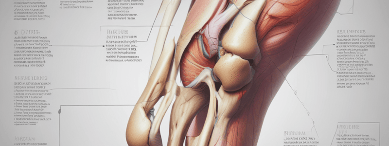
Thigh Innervation
- Obturator nerve (L2-L4) innervates medial thigh
- Tibial nerve (L4-S3) innervates posterior thigh and leg, plantar foot
- Femoral nerve (L2-L4) innervates anterior thigh
- Pudendal nerve (S2-S4) supplies perineum and travels with internal pudendal artery
- Sciatic nerve (L4-S3) travels in posterior compartment and divides into tibial and common peroneal nerves
- Lumbosacral plexus provides motor innervation to the thigh and leg
Motor Innervation
- Lumbar plexus (T12-L4) provides motor innervation to the thigh
- Femoral nerve (L2-L4) provides motor innervation to the anterior thigh, including pectineus and iliopsoas
- Obturator nerve (L2-L4) provides motor innervation to the medial thigh, including obturator externus and adductor magnus
- Sacral plexus (L4-S4) provides motor innervation to the posterior thigh and leg, including gluteal muscles and hamstring muscles
- Sciatic nerve (L4-S3) provides motor innervation to the posterior thigh and leg, including hamstring muscles and gastrocnemius
Sensory Innervation
- Anterior division of lumbar plexus provides sensory innervation to the hip joint and anterior thigh
- Posterior division of lumbar plexus provides sensory innervation to the posterior thigh and leg
- Obturator nerve (L2-L4) provides sensory innervation to the medial thigh and hip joint
- Femoral nerve (L2-L4) provides sensory innervation to the anterior thigh and hip joint
- Posterior femoral cutaneous nerve provides sensory innervation to the posterior thigh and gluteal region
- Pudendal nerve (S2-S4) provides sensory innervation to the perineum and genital area
Hip Joint
- Articular surfaces of the hip joint include the lunate surface of the acetabulum and the head of the femur
- Intrinsic ligaments of the hip joint include the ligament of the head of the femur, transverse acetabular ligament, and fibrous capsule
- Blood supply to the head and neck of the femur is provided by the retinacular arteries, which are branches of the obturator artery
- Fractures of the neck of the femur can lead to avascular necrosis of the femoral head
Thigh Muscles
- Anterior compartment of the thigh includes the quadriceps muscles (vastus lateralis, vastus intermedius, rectus femoris, and vastus medialis)
- Medial compartment of the thigh includes the adductor muscles (adductor longus, adductor brevis, and adductor magnus)
- Posterior compartment of the thigh includes the hamstring muscles (biceps femoris, semimembranosus, and semitendinosus)
- Intermuscular septa separate the compartments of the thigh and attach to the femur and linea aspera
Long Bones
- Long bones, such as the femur, are formed through endochondral ossification
- The diaphysis of a long bone is the middle part of the bone and is the site of primary ossification
- The epiphyses of a long bone are the ends of the bone and are the sites of secondary ossification
- The growth plate, or physis, is the cartilaginous area between the diaphysis and epiphysis where bone growth occurs
Femur
- The femur is the longest, strongest bone in the body
- The proximal end of the femur includes the head, neck, and greater and lesser trochanters
- The distal end of the femur includes the medial and lateral femoral condyles and the intercondylar fossa
- The femur has a pectineal line, spiral line, and linea aspera, which are attachment sites for muscles
- The femur ossifies through a primary ossification center in the diaphysis and secondary ossification centers in the epiphyses
Studying That Suits You
Use AI to generate personalized quizzes and flashcards to suit your learning preferences.


