Podcast
Questions and Answers
What is the name of the valve located between the right atrium and right ventricle?
What is the name of the valve located between the right atrium and right ventricle?
What is the name of the valve located between the coronary sinus and the right atrium?
What is the name of the valve located between the coronary sinus and the right atrium?
What type of ventricular muscle is attached to the ventricular wall at two ends?
What type of ventricular muscle is attached to the ventricular wall at two ends?
What is the name of the structure that passes into the cavity of the ventricle?
What is the name of the structure that passes into the cavity of the ventricle?
Signup and view all the answers
What is the name of the structure located on the lateral wall of the right atrium?
What is the name of the structure located on the lateral wall of the right atrium?
Signup and view all the answers
How many openings of pulmonary veins are present in the left atrium?
How many openings of pulmonary veins are present in the left atrium?
Signup and view all the answers
What layer of the heart contains coronary vessels?
What layer of the heart contains coronary vessels?
Signup and view all the answers
What is the function of the sinoatrial node?
What is the function of the sinoatrial node?
Signup and view all the answers
Which valve is located between the left atrium and left ventricle?
Which valve is located between the left atrium and left ventricle?
Signup and view all the answers
What is the function of the coronary arteries?
What is the function of the coronary arteries?
Signup and view all the answers
Which part of the heart is thicker, the right or left ventricle?
Which part of the heart is thicker, the right or left ventricle?
Signup and view all the answers
What is the function of the cardiac conduction system?
What is the function of the cardiac conduction system?
Signup and view all the answers
Which layer of the heart is in contact with blood flow?
Which layer of the heart is in contact with blood flow?
Signup and view all the answers
What is the function of the tricuspid valve?
What is the function of the tricuspid valve?
Signup and view all the answers
What is the weight of the heart in males?
What is the weight of the heart in males?
Signup and view all the answers
What is the location of the Thymus?
What is the location of the Thymus?
Signup and view all the answers
What forms most of the anterior surface of the heart?
What forms most of the anterior surface of the heart?
Signup and view all the answers
What is the shape of the heart?
What is the shape of the heart?
Signup and view all the answers
What is the location of the base of the heart?
What is the location of the base of the heart?
Signup and view all the answers
What is the surface marking of the right border of the heart?
What is the surface marking of the right border of the heart?
Signup and view all the answers
What is the location of the apex of the heart?
What is the location of the apex of the heart?
Signup and view all the answers
What is the number of chambers in the heart?
What is the number of chambers in the heart?
Signup and view all the answers
What is the function of the atrioventricular node in the heart?
What is the function of the atrioventricular node in the heart?
Signup and view all the answers
What is the Bundle of His?
What is the Bundle of His?
Signup and view all the answers
What is the function of the pericardium?
What is the function of the pericardium?
Signup and view all the answers
What is the purpose of the serous pericardium?
What is the purpose of the serous pericardium?
Signup and view all the answers
What is the function of the visceral layer of the serous pericardium?
What is the function of the visceral layer of the serous pericardium?
Signup and view all the answers
What is the pericardial cavity filled with?
What is the pericardial cavity filled with?
Signup and view all the answers
What is the location of the atrioventricular node?
What is the location of the atrioventricular node?
Signup and view all the answers
What is the significance of the patient's symptoms in the case presentation?
What is the significance of the patient's symptoms in the case presentation?
Signup and view all the answers
What is the normal pulse rate for an adult?
What is the normal pulse rate for an adult?
Signup and view all the answers
What is the normal fetal pulse rate?
What is the normal fetal pulse rate?
Signup and view all the answers
What is the condition of the mitral valve revealed by echocardiography?
What is the condition of the mitral valve revealed by echocardiography?
Signup and view all the answers
What is the condition of the left atrium according to the echocardiography results?
What is the condition of the left atrium according to the echocardiography results?
Signup and view all the answers
What is the type of heart rhythm disorder revealed by the electrocardiogram?
What is the type of heart rhythm disorder revealed by the electrocardiogram?
Signup and view all the answers
What is a potential complication of the ischemic cascade?
What is a potential complication of the ischemic cascade?
Signup and view all the answers
What is a characteristic of modifiable risk factors for cardiac problems?
What is a characteristic of modifiable risk factors for cardiac problems?
Signup and view all the answers
What is the result of heart cells dying due to a blocked coronary artery?
What is the result of heart cells dying due to a blocked coronary artery?
Signup and view all the answers
Study Notes
Cardiovascular System
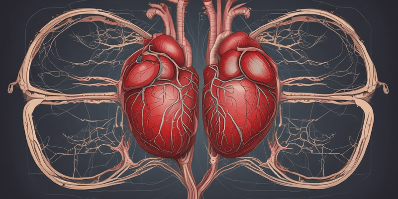
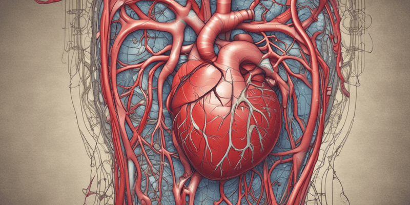
- The cardiovascular system consists of the heart, vessels, and blood.
Heart
- The heart is cone-shaped, weighing approximately 320 grams in males and 270 grams in females.
- It has three surfaces: anterior (sternocostal), inferior (diaphragmatic), and left (pulmonary).
- The heart is located in the middle mediastinum, with one-third on the right side and two-thirds on the left side of the midline.
- The apex of the heart is below and to the left, related to the 5th left intercostal space, 8 cm from the midline.
Position of the Heart
- The base of the heart is located in the superior and posterior regions.
- The apex is inferior and anterior.
Chambers of the Heart
- The heart has four chambers: two atria and two ventricles.
- The right ventricle forms most of the anterior surface.
Grooves on the Heart
- There are two grooves on the anterior surface: the coronary sulcus (or atrioventricular sulcus) and the anterior interventricular sulcus.
- There are two grooves on the inferior surface: the coronary sulcus (or atrioventricular sulcus) and the inferior interventricular sulcus.
Surface Marking of the Heart
- The right border of the heart extends from the 3rd right costal cartilage to the 6th costal cartilage, 1 cm to the right of the sternal border.
- The left border extends from the 2nd left costal cartilage to the 5th left intercostal space, 8 cm from the midline.
Valves of the Heart
- There are four valves: tricuspid valve, mitral valve, pulmonary valve, and aortic valve.
Right Atrium
- The posterior smooth part is the sinus venarum.
- The anterior part has pectinate muscles.
- There is a crista terminalis on the lateral wall.
- The fossa ovalis is on the medial wall.
- The valve of inferior vena cava (Ostashian valve) and the valve of coronary sinus (Thebesian valve) are present.
Left Atrium
- The wall is smooth, except in the left auricle.
- There are four openings of pulmonary veins.
- The mitral valve is between the left atrium and left ventricle.
Ventricular Muscles
- There are three types of ventricular muscles: ridge, bridge, and papillary.
Layers of the Heart
- There are three layers: epicardium (outer layer), myocardium (thick middle layer), and endocardium (inner layer).
Coronary Arteries
- The coronary arteries originate from the aorta and supply the heart with blood flow.
- The main coronary artery lies on the surface of the heart (epicardial coronary arteries).
- Small penetrating arteries supply the myocardial muscle.
Cardiac Conduction System
- The cardiac conduction system controls the heart rate.
- The sinoatrial node (SA node) initiates impulses (70-80 times per minute) and is located in the back wall of the right atrium near the entrance of the vena cava superior.
- The atrioventricular node (AV node) is located in the bottom of the right atrium near the septum and conducts impulses more slowly, causing a delay.
Pericardium
- The pericardium is a double-walled sac around the heart.
- It is composed of the fibrous pericardium and the serous pericardium.
- The pericardium protects and anchors the heart, prevents overfilling, and allows for a friction-free environment.
Clinical Findings
- Patient presentation: shortness of breath, fatigue, and racing heart during exercise.
- Physical examination: pulse rate of 122 bpm, auscultation reveals a mid-diastolic murmur and systolic "snap" over the heart apex, and crackles detected in all lung lobes.
- Echocardiography reveals calcification in both leaflets of the mitral valve, doming of the anterior leaflet, and thickened and shortened chordae tendineae.
Main Contributing Risk Factors to Cardiac Problems
- Hypertension
- Hyperlipidemia
- Smoking
- Diabetes
These are considered modifiable risk factors.
Studying That Suits You
Use AI to generate personalized quizzes and flashcards to suit your learning preferences.
Description
This quiz covers the components of the cardiovascular system, including the heart, vessels, blood, and their functions. It also covers the location of the heart in the mediastinum and the thymus.


