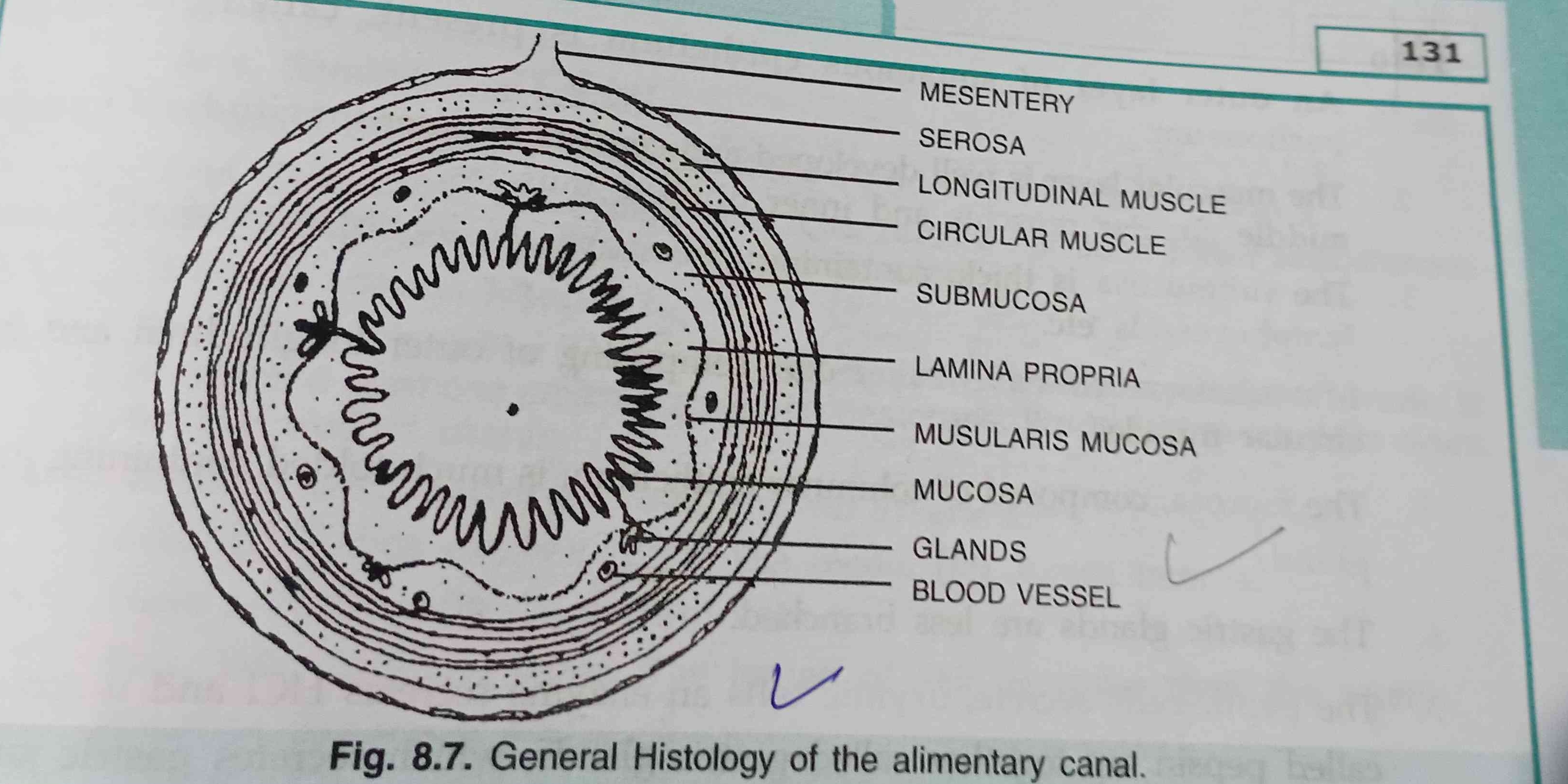
What are the various parts labeled in the histology diagram of the alimentary canal?

Understand the Problem
The question seems to involve the identification and understanding of the components of a histological diagram of the alimentary canal, as labeled in the provided image.
Answer
Mesentery, serosa, longitudinal muscle, circular muscle, submucosa, lamina propria, muscularis mucosa, mucosa, glands, blood vessel.
The labeled parts in the histology diagram of the alimentary canal include: mesentery, serosa, longitudinal muscle, circular muscle, submucosa, lamina propria, muscularis mucosa, mucosa, glands, and blood vessel.
Answer for screen readers
The labeled parts in the histology diagram of the alimentary canal include: mesentery, serosa, longitudinal muscle, circular muscle, submucosa, lamina propria, muscularis mucosa, mucosa, glands, and blood vessel.
More Information
The diagram illustrates the basic layers and structures found in the alimentary canal, essential for digestion and absorption.
Tips
A common mistake is confusing the order of the muscular layers. Remember, the longitudinal muscle is typically outer to the circular muscle.
Sources
- Layers of the Alimentary Canal – Boundless Anatomy and Physiology - university.pressbooks.pub
AI-generated content may contain errors. Please verify critical information