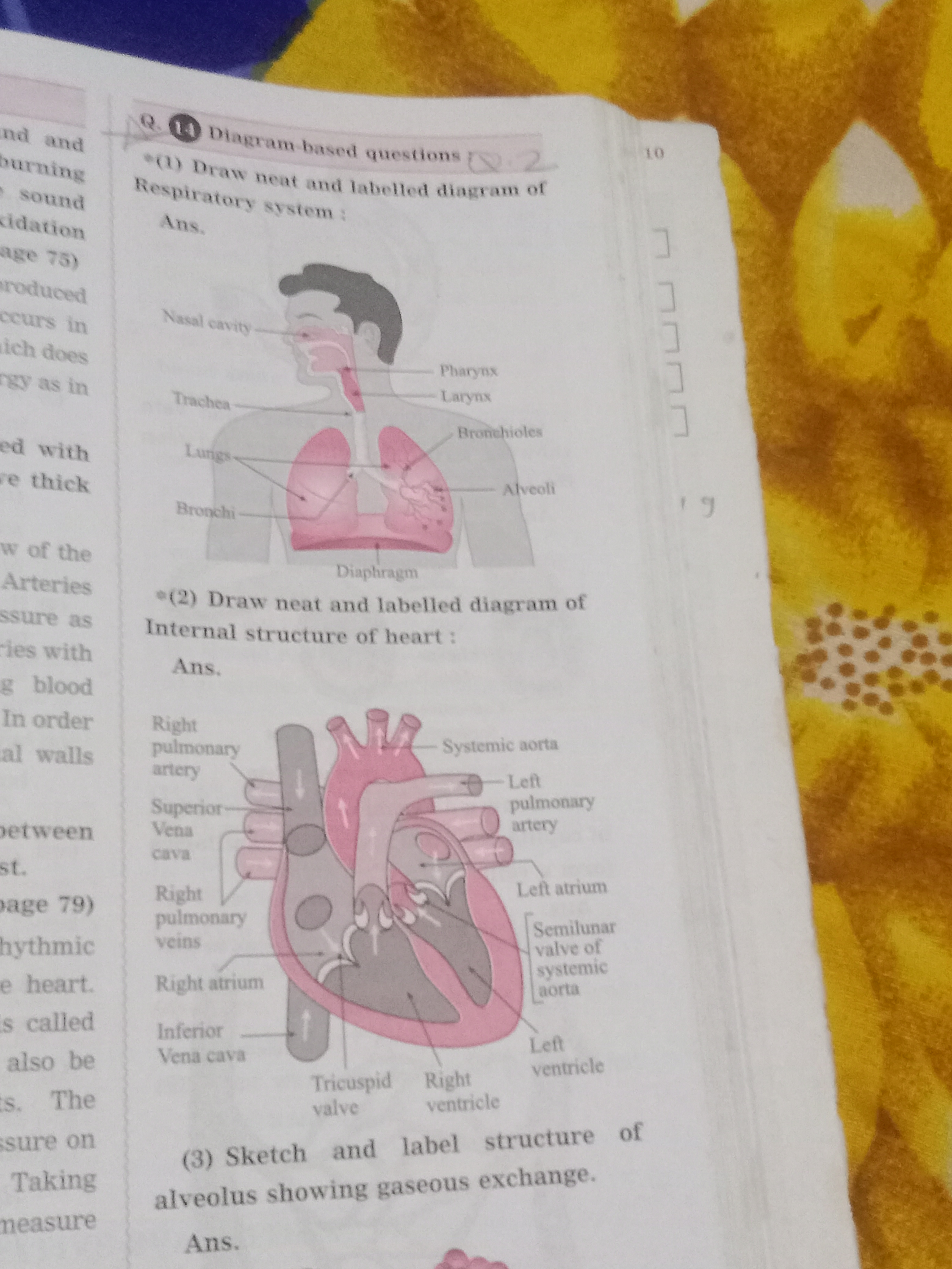
Draw neat and labelled diagram of respiratory system and internal structure of heart. Sketch and label structure of alveolus showing gaseous exchange.

Understand the Problem
The question is asking for the drawing and labeling of diagrams related to the respiratory system and the heart. The focus is on understanding and illustrating the anatomical structures relevant to these systems.
Answer
The image has detailed, labeled diagrams of the respiratory system, heart, and alveolus structure.
The image shows neat and labeled diagrams of the respiratory system, the internal structure of the heart, and the alveolus structure. These diagrams help illustrate how the respiratory and circulatory systems work, focusing on gas exchange in the alveoli and the heart's anatomy.
Answer for screen readers
The image shows neat and labeled diagrams of the respiratory system, the internal structure of the heart, and the alveolus structure. These diagrams help illustrate how the respiratory and circulatory systems work, focusing on gas exchange in the alveoli and the heart's anatomy.
More Information
Diagrams enhance understanding of complex biological systems like the respiratory and circulatory systems by visualizing how different parts contribute to essential functions such as oxygen transport and gas exchange.
Tips
Ensure the diagrams are clear and labels are legible to aid in learning.
Sources
- Organs and Structures of the Respiratory System - Lumen Learning - courses.lumenlearning.com
- Draw neat and labeled diagrams. a. Respiratory system b. Internal ... - byjus.com
- Fully labelled diagram of the alveolus in the lungs showing gaseous ... - stock.adobe.com
AI-generated content may contain errors. Please verify critical information