Podcast
Questions and Answers
What is the primary function of veins?
What is the primary function of veins?
Veins conduct blood from the capillaries toward the heart.
Veins in the systemic circuit carry oxygen-rich blood.
Veins in the systemic circuit carry oxygen-rich blood.
False (B)
What are the smallest veins called, and what is their approximate diameter?
What are the smallest veins called, and what is their approximate diameter?
The smallest veins are called venules, with a diameter of 8-100 µm.
Which type of venule functions similarly to capillaries, especially during inflammatory responses?
Which type of venule functions similarly to capillaries, especially during inflammatory responses?
Approximately what percentage of the body's blood is typically held within the veins at any given time?
Approximately what percentage of the body's blood is typically held within the veins at any given time?
Compare the relative thickness of the tunica media and tunica externa in veins versus arteries.
Compare the relative thickness of the tunica media and tunica externa in veins versus arteries.
Veins generally have thicker walls and more elastin than arteries of comparable size.
Veins generally have thicker walls and more elastin than arteries of comparable size.
Why is blood pressure significantly lower in veins compared to arteries?
Why is blood pressure significantly lower in veins compared to arteries?
How many major veins enter the right atrium of the heart, and what are their names?
How many major veins enter the right atrium of the heart, and what are their names?
Where are superficial veins typically found, and what is their clinical importance?
Where are superficial veins typically found, and what is their clinical importance?
What is a venous plexus?
What is a venous plexus?
Into which unique structures do veins from the brain drain?
Into which unique structures do veins from the brain drain?
The superior vena cava receives systemic blood from all body regions ____ to the diaphragm (excluding the heart wall).
The superior vena cava receives systemic blood from all body regions ____ to the diaphragm (excluding the heart wall).
The inferior vena cava returns blood to the heart from all body regions _____ to the diaphragm.
The inferior vena cava returns blood to the heart from all body regions _____ to the diaphragm.
The superior vena cava is formed by the union of which two large veins?
The superior vena cava is formed by the union of which two large veins?
The inferior vena cava begins inferiorly at the union of which two veins?
The inferior vena cava begins inferiorly at the union of which two veins?
What is the primary function of the hepatic portal system?
What is the primary function of the hepatic portal system?
The hepatic portal system is a series of vessels where two separate _____ beds lie between the arterial supply and the final venous drainage.
The hepatic portal system is a series of vessels where two separate _____ beds lie between the arterial supply and the final venous drainage.
Blood exits the liver sinusoids and eventually returns to the inferior vena cava via which veins?
Blood exits the liver sinusoids and eventually returns to the inferior vena cava via which veins?
Distinguish between the hepatic veins and the hepatic portal vein regarding blood flow direction relative to the liver.
Distinguish between the hepatic veins and the hepatic portal vein regarding blood flow direction relative to the liver.
Which major tributary of the hepatic portal vein drains the small intestine and the ascending and transverse colon?
Which major tributary of the hepatic portal vein drains the small intestine and the ascending and transverse colon?
Which vein unites with the superior mesenteric vein to form the hepatic portal vein?
Which vein unites with the superior mesenteric vein to form the hepatic portal vein?
The inferior mesenteric vein primarily drains which parts of the large intestine?
The inferior mesenteric vein primarily drains which parts of the large intestine?
What condition, often caused by cirrhosis, leads to increased blood pressure in the hepatic portal system?
What condition, often caused by cirrhosis, leads to increased blood pressure in the hepatic portal system?
What is the function of portal-systemic (portal-caval) anastomoses?
What is the function of portal-systemic (portal-caval) anastomoses?
List the three main locations where portal-systemic anastomoses occur.
List the three main locations where portal-systemic anastomoses occur.
What is 'caput medusae'?
What is 'caput medusae'?
Flashcards
What are veins?
What are veins?
Blood vessels conducting blood from capillaries to the heart.
What are venules?
What are venules?
The smallest veins, functioning similarly to capillaries.
What is the hepatic portal system?
What is the hepatic portal system?
A system of veins returning blood from the digestive organs to the liver for processing before re-entering general circulation.
What are the venae cavae?
What are the venae cavae?
Signup and view all the flashcards
What is the superior vena cava?
What is the superior vena cava?
Signup and view all the flashcards
What is the inferior vena cava?
What is the inferior vena cava?
Signup and view all the flashcards
What are the major tributaries of the hepatic portal vein?
What are the major tributaries of the hepatic portal vein?
Signup and view all the flashcards
What is portal hypertension?
What is portal hypertension?
Signup and view all the flashcards
What are Portal-Systemic Anastomoses?
What are Portal-Systemic Anastomoses?
Signup and view all the flashcards
What vein drains the small intestine and part of the large intestine?
What vein drains the small intestine and part of the large intestine?
Signup and view all the flashcards
Which vein drains the distal region of the colon and the superior rectum?
Which vein drains the distal region of the colon and the superior rectum?
Signup and view all the flashcards
What is caput medusae?
What is caput medusae?
Signup and view all the flashcards
What is the difference in the tunica composition of veins and arteries?
What is the difference in the tunica composition of veins and arteries?
Signup and view all the flashcards
Lumen Differences for Veins and Arteries
Lumen Differences for Veins and Arteries
Signup and view all the flashcards
Study Notes
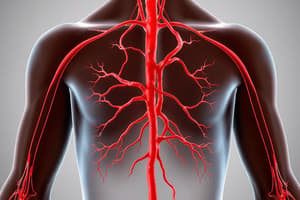
Venous Vessels
- Veins carry blood from capillaries to the heart
- Systemic circuit veins carry relatively oxygen-poor blood, except for pulmonary veins, which carry oxygen-rich blood from the lungs
- Venules, the smallest veins, measure 89-100 µm in diameter
- Postcapillary venules consist of an endothelium with pericytes and function like capillaries
- During inflammation, postcapillary venules allow more fluid and leukocytes to leave the circulation than capillaries
- Larger venules have a tunica media with one or two smooth muscle layers and a thin tunica externa
Veins vs Arteries
- Veins form when venules join
- Veins differ structurally from arteries
- Veins have a larger lumen than comparable arteries, holding 65% of the body's blood at any given time
- The tunica externa is thicker than the tunica media in veins, which is the opposite of arteries
- Longitudinal bands of smooth muscle further thicken the tunica externa in the venae cavae
- Veins have less elastin in their walls and don't need to dampen pulsations like arteries do
- Vein walls are thinner than comparable arteries
- Blood pressure is lower in veins due to the pressure drop in arterioles and capillary beds
Systemic Veins
- While veins typically run with corresponding arteries, their distributions differ
- While one large systemic artery (aorta) exits the left ventricle, three major veins enter the heart's right atrium: the superior and inferior venae cavae and the coronary sinus
- Large and medium-sized arteries are deep and accompanied by deep veins of the same name, as well as superficial veins found just beneath the skin
- Superficial veins are important for drawing blood or placing intravenous lines, but are susceptible to cuts or injuries
Venous Patterns and Drainage
- Two or more parallel veins often drain a body region instead of a single larger vein
- Multiple veins can anastomose to form a venous plexus
- The brain and digestive tract have unique venous drainage patterns
- Brain veins drain into dural venous sinuses, which are endothelium-lined channels supported by dura mater
- Venous blood from digestive organs enters the hepatic portal system, passing through liver capillaries before reentering general systemic circulation
Venae Cavae and Tributaries
-
Venae Cavae refers to the unpaired superior and inferior venae cavae
-
These are the body's two largest veins, emptying directly into the right atrium of the heart
-
The name "vena cava" means "cavelike vein"
-
Superior Vena Cava receives systemic blood from body regions superior to the diaphragm, excluding the heart wall
-
It arises from the union of the left and right brachiocephalic veins behind the manubrium and descends to the right atrium
-
The left brachiocephalic vein is longer and more horizontal than the right, which is more vertical
-
Each brachiocephalic vein forms from the union of an internal jugular vein and a subclavian vein
-
Inferior Vena Cava ascends along the posterior abdominal wall and is the widest vessel
-
It returns blood to the heart from regions inferior to the diaphragm
-
It begins inferiorly at the union of the two common iliac veins on vertebra L5
-
Ascends on the right side of the vertebral bodies, to the right of the abdominal aorta
-
Penetrates the central tendon of the diaphragm at T8 before joining the right atrium
Hepatic Portal System
- The hepatic portal system is a specialized vascular circuit for digestion
- It picks up nutrients from the stomach and intestines, delivering them to the liver for processing and storage
- It is a series of vessels with two capillary beds between arterial supply and venous drainage
- Capillaries in the stomach and intestines receive digested nutrients, draining into tributaries of the hepatic portal vein
- The hepatic portal vein delivers nutrient-rich blood to liver sinusoids
- Nutrients reach liver cells, and toxins from the digestive tract are broken down
- Blood then enters hepatic veins and the inferior vena cava, reentering systemic circulation
Hepatic Portal Vein Tributaries
- The hepatic portal system includes multiple tributaries
- The superior mesenteric vein ascends to the right of the superior mesenteric artery
- It drains the entire small intestine, the first half of the large intestine (ascending and transverse colon), and part of the stomach
- Its superior part lies behind the stomach and pancreas
- The splenic vein drains venous blood from the spleen, running horizontally behind the stomach and pancreas
- It joins the superior mesenteric vein to form the hepatic portal vein
- Tributaries from the splenic artery correspond to the splenic vein tributaries
- This process carries spleen microbes to the liver for destruction after immune responses
- The inferior mesenteric vein ascends on the posterior abdominal wall, left of the inferior mesenteric artery
- It drains the distal colon region and the superior rectum, emptying into the splenic vein behind the stomach and pancreas
Hepatic Portal Vein Anatomy
- The hepatic portal vein is a short, vertical vessel inferior to the liver and anterior to the inferior vena cava
- Inferiorly, it starts behind the pancreas from the union of the superior mesenteric and splenic veins
- Superiorly, it enters the liver's underside, dividing into right and left branches that reach liver sinusoids
- The hepatic portal vein also receives the right and left gastric veins from the stomach
Portal Systemic Anastomoses
-
Liver cirrhosis from conditions like chronic alcoholism, blocks blood flow through liver sinusoids, raising pressure in the hepatic portal system, leading to portal hypertension
-
Portal system veins form anastomoses veins that drain into the venae cavae in order to provide alternate routes for blood to return to the heart
-
The main anastomoses are
-
Veins in the inferior esophagus
-
Hemorrhoidal veins in the anal canal wall
-
Superficial veins in the abdominal wall around the navel
-
Connecting veins are small and can swell/burst when forced to carry large blood volumes
-
In cirrhosis patients, esophageal veins can tear and cause them to vomit blood, hemorrhoids can swell up, and snakelike and distended veins can show through the skin around the navel
-
Ruptured esophageal veins can cause mortality in 50% of cases and an additional 30% with recurrence
-
Treatment involves injecting a hardening agent/tying them off or implantation of a tube from the inferior vena cava behind the liver to the portal vein to create a new portalcaval shunt, to relieve hypertension
Studying That Suits You
Use AI to generate personalized quizzes and flashcards to suit your learning preferences.