Podcast
Questions and Answers
What is the primary location of the stomach within the abdominal cavity?
What is the primary location of the stomach within the abdominal cavity?
- Left side, caudal to the diaphragm (correct)
- Right side, adjacent to the intestines
- Right side, below the liver
- Middle, above the pancreas
Which of the following types of tumors is a benign condition of the stomach?
Which of the following types of tumors is a benign condition of the stomach?
- Gastric adenocarcinoma
- Gastric lymphoma
- Gastric leiomyoma (correct)
- Gastrointestinal stromal tumor
Which radiographic method is considered the most sensitive for diagnosing gastric cancer?
Which radiographic method is considered the most sensitive for diagnosing gastric cancer?
- CT scan
- Endoscopy (correct)
- Ultrasound
- MRI
What characteristic feature is diagnostic for gastric lipomas on CT?
What characteristic feature is diagnostic for gastric lipomas on CT?
Which of the following is NOT classified as a malignant condition related to the stomach?
Which of the following is NOT classified as a malignant condition related to the stomach?
What is the typical feature of gastric leiomyomas on radiographic imaging?
What is the typical feature of gastric leiomyomas on radiographic imaging?
What type of gastritis is specifically mentioned as often related to infections?
What type of gastritis is specifically mentioned as often related to infections?
Which condition is characterized as a congenital gastric anomaly?
Which condition is characterized as a congenital gastric anomaly?
What is the primary use of CT scans in relation to gastric carcinoma?
What is the primary use of CT scans in relation to gastric carcinoma?
Which characteristic is associated with gastric lymphoma on a CT scan?
Which characteristic is associated with gastric lymphoma on a CT scan?
What percentage of gastric cancers are represented by ulcerated carcinomas?
What percentage of gastric cancers are represented by ulcerated carcinomas?
In which situation is ultrasound most likely to be useful for gastric diagnosis?
In which situation is ultrasound most likely to be useful for gastric diagnosis?
What radiographic feature may indicate gastritis on a CT scan?
What radiographic feature may indicate gastritis on a CT scan?
What is the role of fluoroscopy in the diagnosis of gastric carcinoma?
What is the role of fluoroscopy in the diagnosis of gastric carcinoma?
What does gastritis refer to?
What does gastritis refer to?
What is the typically observed characteristic of gastric lymphoma in terms of tumor appearance?
What is the typically observed characteristic of gastric lymphoma in terms of tumor appearance?
What is the primary anatomical feature that distinguishes the small intestine?
What is the primary anatomical feature that distinguishes the small intestine?
Which of the following is NOT a functional unit of the small intestine?
Which of the following is NOT a functional unit of the small intestine?
What percentage of mechanical intestinal obstructions are due to small bowel obstruction?
What percentage of mechanical intestinal obstructions are due to small bowel obstruction?
Which imaging technique is more sensitive for diagnosing small bowel obstruction?
Which imaging technique is more sensitive for diagnosing small bowel obstruction?
What is a common feature seen on an abdominal radiograph indicating small bowel obstruction?
What is a common feature seen on an abdominal radiograph indicating small bowel obstruction?
What does the small bowel feces sign indicate when observed on CT?
What does the small bowel feces sign indicate when observed on CT?
Which carcinoma is significantly less common in the small bowel compared to the colon?
Which carcinoma is significantly less common in the small bowel compared to the colon?
What characteristic feature of small bowel ischemia can be observed on a CT scan?
What characteristic feature of small bowel ischemia can be observed on a CT scan?
Flashcards are hidden until you start studying
Study Notes
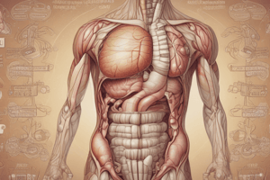
Stomach
- Muscular organ located in the upper abdomen, between the esophagus and duodenum, primarily on the left side, at T10 level from diaphragm.
Radiographic Features
- Outlined on fluoroscopy during imaging studies.
Related Pathology
-
Neoplastic
- Benign:
- Gastric leiomyoma: Rare, asymptomatic, usually found incidentally.
- Gastric lipoma: Rare benign mesenchymal tumor.
- Malignant:
- Gastric adenocarcinoma: Most common gastric malignancy, third most common GI cancer.
- Gastric lymphoma: Can be primary or secondary.
- Gastrointestinal stromal tumor.
- Benign:
-
Inflammatory:
- Gastritis: Inflammation of gastric mucosa, can be acute or chronic.
-
Infections:
- Phlegmonous gastritis: Severe infection causing inflammation.
-
Congenital:
- Gastric duplication cyst: Rare congenital anomaly.
-
Other Conditions:
- Gastric volvulus: Twisting of stomach, leading to obstruction.
- Gastric varix: Dilated veins in stomach walls.
- Gastric diverticulum: Outpouching in the gastric wall.
- Gastric emphysema: Presence of gas in gastric wall.
Gastric Leiomyomas
- Presents as well-defined solid mass with low homogeneous contrast enhancement.
- Calcifications and intratumoral hemorrhages are rare.
Gastric Lipoma
- Diagnosed on CT as well-defined fat-density lesions (-70 to -120 HU).
Gastric Adenocarcinoma
- Most common type of gastric cancer, arising from gastric epithelium.
- Endoscopy is the gold standard for diagnosis, visualizing tumor location and involvement.
- Radiographic features include polypoid masses or ulcers.
- Staging primarily done through CT for tumor spread, nodal involvement, and metastases.
Gastric Lymphoma
- Can represent secondary systemic involvement or primary gastric malignancy.
- CT typically shows thickened stomach wall (2-4 cm) with lateral tumor extension and possible necrosis.
Gastritis
- Characterized by mucosal inflammation, detectable by thickened gastric folds on CT.
Small Bowel
- Section of bowel located between the stomach and colon, includes duodenum, jejunum, and ileum.
Related Pathology
- Conditions include small bowel obstruction, ischemia, and malignancies like adenocarcinoma and lymphoma.
Small Bowel Obstruction (SBO)
- Accounts for 80% of mechanical intestinal obstructions, with ~5% mortality.
- Abdominal radiograph: 50-60% sensitivity, shows dilated loops proximal to obstruction and specific gas-fluid levels.
- CT: More sensitive, revealing dilated loops (>2.5 cm), with normal or collapsed loops distally.
Small Bowel Ischemia
- Life-threatening condition due to blood flow disturbance to the bowel wall.
Adenocarcinoma of the Small Bowel
- Much rarer than colorectal cancer; CT shows soft-tissue mass with heterogeneous attenuation, often with annular narrowing and irregular edges.
Studying That Suits You
Use AI to generate personalized quizzes and flashcards to suit your learning preferences.