Podcast
Questions and Answers
Which nutrient deficiency is most commonly associated with macrocytic states?
Which nutrient deficiency is most commonly associated with macrocytic states?
- Vitamin C
- Iron
- Folic acid and Vitamin B12 (correct)
- Vitamin D
What characteristic distinguishes macrocytic red blood cells?
What characteristic distinguishes macrocytic red blood cells?
- They are smaller than normal cells.
- They have a higher MCHC than normal cells.
- They are non-nucleated and have a diameter greater than 8 microns. (correct)
- They contain a central area of redness.
What condition is likely to cause a red blood cell not to mature effectively?
What condition is likely to cause a red blood cell not to mature effectively?
- Excessive hydration
- Nutritional deficiency (correct)
- Dehydration
- High oxygen levels
What is the primary function of hemoglobin in red blood cells?
What is the primary function of hemoglobin in red blood cells?
What does anisochromia indicate about red blood cells?
What does anisochromia indicate about red blood cells?
Which of the following factors contributes to the red cell membrane's integrity?
Which of the following factors contributes to the red cell membrane's integrity?
What does MCH stand for in hematology?
What does MCH stand for in hematology?
How does glucose enter red blood cells for energy?
How does glucose enter red blood cells for energy?
What are blister cells primarily associated with?
What are blister cells primarily associated with?
What causes the formation of Degmacytes or bite cells?
What causes the formation of Degmacytes or bite cells?
Which type of ribosomal stippling is associated with lead poisoning?
Which type of ribosomal stippling is associated with lead poisoning?
What is the characteristic appearance of Heinz bodies?
What is the characteristic appearance of Heinz bodies?
Which stain is used to confirm the presence of Howell-Jolly bodies?
Which stain is used to confirm the presence of Howell-Jolly bodies?
What does the presence of ringed sideroblasts indicate?
What does the presence of ringed sideroblasts indicate?
What is the main feature of Hb CC crystals?
What is the main feature of Hb CC crystals?
Which type of parasites is associated with a Maltese cross formation?
Which type of parasites is associated with a Maltese cross formation?
What causes the formation of Pappenheimer bodies?
What causes the formation of Pappenheimer bodies?
What does the Rouleaux formation indicate about the blood?
What does the Rouleaux formation indicate about the blood?
What are Heinz bodies primarily associated with?
What are Heinz bodies primarily associated with?
What is a common appearance of Dacrocytes?
What is a common appearance of Dacrocytes?
How are Schistocytes formed?
How are Schistocytes formed?
What does the presence of agglutination in blood indicate?
What does the presence of agglutination in blood indicate?
What is the characteristic feature of hypochromic red blood cells?
What is the characteristic feature of hypochromic red blood cells?
Which condition is most likely associated with microcytic red blood cells?
Which condition is most likely associated with microcytic red blood cells?
What grading corresponds to a central pallor that is two-thirds of a red blood cell's diameter in hypochromasia?
What grading corresponds to a central pallor that is two-thirds of a red blood cell's diameter in hypochromasia?
Which of the following describes acanthocytes?
Which of the following describes acanthocytes?
What defines a normocytic red blood cell?
What defines a normocytic red blood cell?
Which of the following best describes spherocytes?
Which of the following best describes spherocytes?
Which type of macrocyte has no central area of pallor and is associated with megaloblastic anemia?
Which type of macrocyte has no central area of pallor and is associated with megaloblastic anemia?
What is the primary cause of schistocytes?
What is the primary cause of schistocytes?
Which condition is associated with echinocytes?
Which condition is associated with echinocytes?
Stomatocytes are characterized by which shape?
Stomatocytes are characterized by which shape?
Drepanocytes are associated with which abnormal condition?
Drepanocytes are associated with which abnormal condition?
What is a common feature of target cells (codocytes)?
What is a common feature of target cells (codocytes)?
What condition is associated with polychromasia?
What condition is associated with polychromasia?
Echinocytes are also known as what?
Echinocytes are also known as what?
Study Notes
Red Blood Cell Morphology
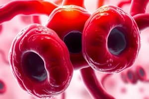
- Red blood cells (RBCs) are non-nucleated, biconcave disc-like cells with a diameter of 6-8 microns.
- They have a central area of pallor that represents about 1/3 of the cell's diameter, providing extra surface area.
- RBCs require a healthy cell membrane composed of proteins, lipids, and carbohydrates, deformability, sufficient hemoglobin, and a balance between intracellular and extracellular environments.
Red Blood Cell Size (MCV)
- Normocytic: 80-100 fL, normal structure, quantity may be affected.
- Macrocytic: > 100 fL, cells did not mature properly, associated with nutritional deficiencies, liver diseases, or megaloblastic states.
- Microcytic: < 80 fL, smaller in size, low hemoglobin production, associated with conditions like thalassemia and iron deficiency anemia.
Red Blood Cell Color (MCH & MCHC)
- Normochromic: Normal MCH (27-32 pg) and MCHC (32-36%), normal red cell structure.
- Hypochromic: Pale red blood cells, low hemoglobin, central pallor area > 1/3, associated with iron deficiency anemia, thalassemia, and other conditions.
- Hyperchromic: No central pallor area, appears purely red, susceptible to hemolysis, does not mean increased hemoglobin content.
Polychromasia
- Variation in hemoglobin content, resulting in a blue-tinged appearance due to residual RNA.
- Represents reticulocytes, young red blood cells.
- Elevated polychromasia can indicate hemolytic anemia.
Red Blood Cell Shape (Poikilocytosis)
- Poikilocytosis refers to any variation in red blood cell shape.
- Different shapes can arise from developmental abnormalities, membrane abnormalities, trauma, or abnormal hemoglobin content.
Developmental Macrocytosis
- Oval Macrocytes: Elongated with wide diameter, increased MCV, associated with megaloblastic erythropoiesis and megaloblastic anemia.
- Round Hypochromic Macrocyte: Seen in conditions like alcoholism, hypothyroidism, and liver disease.
- Blue-Tinged Macrocyte: Found in neonates and anemic individuals.
Membrane Abnormalities
- Acanthocytes (Thorn Cells): Spheroid with irregular spikes, often found in hemolytic anemia, alcoholism, and other conditions.
- Echinocytes (Sea-Urchin Cells): Crenated RBCs with evenly distributed short projections, can occur in hypertonic solutions or when cells lack energy.
- Stomatocytes (Mouth Cells): Bowl-shaped cells with increased membrane permeability to sodium, often seen in alcoholism, liver diseases, and Rh null conditions.
- Elliptocytes: Elongated with narrow diameter, rod or cigar-shaped, associated with elliptocytosis, iron deficiency anemia, and thalassemia.
- Spherocytes: Hyperchromic and ball-shaped, low surface area to volume ratio, susceptible to hemolysis, seen in hereditary spherocytosis, hemolytic anemia, and other conditions.
Trauma
- Schistocytes: Fragmented RBCs, often arise from trauma in blood vessels caused by clots, damaged vessels, or prosthetics.
- Keratocytes: Schistocytes with horn-like projections.
Abnormal Hb Content
- Drepanocytes (Sickle Cells): Leaf-like or crescent-shaped cells formed due to polymerization of abnormal hemoglobin S.
Other
- Dacryocytes (Tear-drop Cells): Tear-drop shaped, formed due to squeezing and fragmentation in the spleen.
- Codocytes (Target Cells): Cells with a peripheral rim of hemoglobin surrounding a clear space, often seen in thalassemia and liver diseases.### Blister Cells
- They are associated with microangiopathic hemolytic anemia (MAHA)
- They are also known as helmet cells.
- Blister cells are formed due to trauma in red cells.
- They are precursor cells to schistocytes.
Degmacyte
- Also known as bite cells.
- They look like a bitten piece of donut.
- Formed because of Heinz bodies caused by oxidative stress.
- Heinz bodies are formed due to G6PD deficiency.
- The spleen removes Heinz bodies, leaving a permanent mark on the cell.
Basophilic Stipplings
- Evidence of ribosomes and RNA precipitation due to heavy metal poisoning in the erythropoietic cells.
- D-ALA is affected by heavy metal poisoning, which impacts heme synthesis.
- D-ALA helps form heme.
- Accumulation of ribosomes and RNA within the cell.
- Ribosomes and RNA help form globin.
- Basophilic stipplings can have a donut appearance with blueberries on top.
- Two types of basophilic stipplings:
- Fine: precipitation of excessive RNA associated with polychromasia.
- Coarse: problems with hemoglobin synthesis, particularly in cases of lead poisoning.
Cabot Ring
- Result of improper cell development: Microtubules and mitotic spindle remain in the cell.
- Fragments of nuclear material.
Howell-Jolly Bodies
- Small round fragments of the nucleus caused by karyorrhexis, where the nucleus is excluded.
- Staining techniques confirm nuclear remnants.
- Feulgen stain is the most specific stain for Howell-Jolly bodies.
Heinz Bodies
- Formed due to oxidative stress.
- They have a golf ball-like appearance.
- They occur due to defects in the pentose-phosphate pathway or G6PD deficiency, which increases red cell susceptibility to oxidative stress.
- Supravital stain is used to identify Heinz bodies.
Ringed Sideroblast
- An immature cell with an accumulation of iron deposits.
- Sidero means "iron."
- They are present in cases of lead poisoning and porphyria (insufficient porphyrin IX synthesis).
- Prussian blue (Perl's stain) is used to confirm the presence of iron.
Pappenheimer Bodies
- Basophilic inclusions that cluster near the cell's periphery.
- They form due to the accumulation of ribosomes, mitochondria, and unused iron.
Hemoglobin H Inclusion
- Represents the precipitation of abnormal hemoglobin H.
- Occurs in a form of alpha thalassemia accompanied by hemolysis.
- Formed due to abnormalities within the globin chain.
- Supravital stain is used to identify HbH inclusions.
Hb CC Crystals
- Hexagonal hemoglobin with blunt ends, stained dark.
- Formed due to the precipitation of hemoglobin C crystals.
Hb SC Crystals
- A combination of abnormal hemoglobin S and C.
Microorganisms
- Malaria:
- Maltese cross formation.
- Thick-borne.
- Sometimes mistaken for malaria.
Agglutination
- Can be reported.
- Occurs due to antibodies (cold agglutinins, hemolytic anemia, or pneumonia).
Rouleaux
- Stack of coins appearance.
- Can indicate increased protein in the blood.
- Excessive antibodies are present.
- Commonly seen in multiple myeloma.
- Adding NSS (normal saline solution) can separate and disperse red blood cells.
Morphology Grading
- Polychromatophilia
- 1+: 1-5 per field
- 2+: 6-10 per field
- 3+: >10 per field
- Helmet cell, Dacrocyte
- 1+: 1-5 per field
- 2+: 6-10 per field
- 3+: >10 per field
- Spherocyte, Acanthocyte
- 1+: 1-5 per field
- 2+: 6-10 per field
- 3+: >10 per field
- Schistocyte
- 1+: 1-5 per field
- 2+: 6-10 per field
- 3+: >10 per field
- Poikilocytosis
- 1+: 3-10 per field
- 2+: 11-20 per field
- 3+: >20 per field
- Codocyte, Burr cell
- 1+: 3-10 per field
- 2+: 11-20 per field
- 3+: >20 per field
- Stomatocyte, Ovalocyte
- 1+: 3-10 per field
- 2+: 11-20 per field
- 3+: >20 per field
- Elliptocyte
- 1+: 3-10 per field
- 2+: 11-20 per field
- 3+: >20 per field
- Rouleaux
- 1+: 3-4 aggregates
- 2+: 5-10 aggregates
- 3+: Numerous aggregates
- Sickle cells: Positive only
- Basophilic stippling: Positive only
- Pappenheimer bodies: Positive only
- Howell-Jolly bodies: Positive only
Studying That Suits You
Use AI to generate personalized quizzes and flashcards to suit your learning preferences.
Related Documents
Description
Explore the essential characteristics of red blood cells including their size, shape, and color metrics. This quiz covers topics such as normocytic, macrocytic, and microcytic classifications, as well as the implications of each type on health. Test your knowledge on RBC morphology and their physiological significance.


