Podcast
Questions and Answers
What are elliptocytes commonly referred to as?
What are elliptocytes commonly referred to as?
- Ovalocytes (correct)
- Target cells
- Sickle cells
- Ovoid cells
What size do mature red blood cells typically measure?
What size do mature red blood cells typically measure?
- 6-8 micrometers (correct)
- 4-6 micrometers
- 8-10 micrometers
- 10-12 micrometers
Which of the following conditions is associated with codocytes?
Which of the following conditions is associated with codocytes?
- Sickle cell anemia
- Liver disease (correct)
- Iron deficiency anemia
- Polycythemia vera
What term refers to an increased variation in red blood cell size?
What term refers to an increased variation in red blood cell size?
Which statement is true about the shape of mature red blood cells?
Which statement is true about the shape of mature red blood cells?
In what condition might you expect to see megaloblastic anemia?
In what condition might you expect to see megaloblastic anemia?
What is the average thickness of a mature red blood cell?
What is the average thickness of a mature red blood cell?
What appearance do codocytes have?
What appearance do codocytes have?
What is a key characteristic of a spherocyte?
What is a key characteristic of a spherocyte?
Which condition is associated with spherocyte formation?
Which condition is associated with spherocyte formation?
What is a distinctive feature of a dacryocyte?
What is a distinctive feature of a dacryocyte?
In which conditions are dacryocytes commonly seen?
In which conditions are dacryocytes commonly seen?
What is the primary cause of stomatocytes?
What is the primary cause of stomatocytes?
Which of the following inclusions is characterized by aggregated RNA?
Which of the following inclusions is characterized by aggregated RNA?
What stain is used to visualize basophilic stippling?
What stain is used to visualize basophilic stippling?
Which condition is NOT typically associated with stomatocytes?
Which condition is NOT typically associated with stomatocytes?
What do echinocytes resemble due to their regular spicules?
What do echinocytes resemble due to their regular spicules?
Which condition is associated with microcytic red blood cells?
Which condition is associated with microcytic red blood cells?
What structural change characterizes a drepanocyte?
What structural change characterizes a drepanocyte?
Macrocytic red blood cells are larger than normal due to defects in what process?
Macrocytic red blood cells are larger than normal due to defects in what process?
What causes schistocytes to form?
What causes schistocytes to form?
Which of the following conditions does NOT typically present with microcytic anemia?
Which of the following conditions does NOT typically present with microcytic anemia?
What condition is hyperchromasia NOT associated with?
What condition is hyperchromasia NOT associated with?
What is a common cause of the elongated appearance seen in drepanocytes?
What is a common cause of the elongated appearance seen in drepanocytes?
In what condition are megaloblastic anemias primarily classified?
In what condition are megaloblastic anemias primarily classified?
Which of the following best describes Pappenheimer bodies?
Which of the following best describes Pappenheimer bodies?
Nucleated red blood cells indicate which physiological condition?
Nucleated red blood cells indicate which physiological condition?
Which of the following conditions are associated with megaloblastic anemia?
Which of the following conditions are associated with megaloblastic anemia?
Which statement is true regarding the cells involved in hemoglobinopathies?
Which statement is true regarding the cells involved in hemoglobinopathies?
What is the significance of finding macrocytosis along with hyperchromasia?
What is the significance of finding macrocytosis along with hyperchromasia?
Which condition does NOT typically lead to increased erythropoiesis?
Which condition does NOT typically lead to increased erythropoiesis?
Which of the following is a characteristic of spherocytosis?
Which of the following is a characteristic of spherocytosis?
Which type of malaria is associated with Plasmodium falciparum?
Which type of malaria is associated with Plasmodium falciparum?
What feature is characteristic of Plasmodium ovale?
What feature is characteristic of Plasmodium ovale?
In grading of hypochromia, what does a grade of 2+ indicate?
In grading of hypochromia, what does a grade of 2+ indicate?
What condition is associated with hyperplasia of red blood cells?
What condition is associated with hyperplasia of red blood cells?
What percentage of red cells must be polychromatophilic to receive a grade of 3+?
What percentage of red cells must be polychromatophilic to receive a grade of 3+?
What is the significance of Maurer’s dots?
What is the significance of Maurer’s dots?
Which species of Plasmodium is considered benign?
Which species of Plasmodium is considered benign?
What indicates a condition of 4+ in grading hypochromia?
What indicates a condition of 4+ in grading hypochromia?
Flashcards are hidden until you start studying
Study Notes
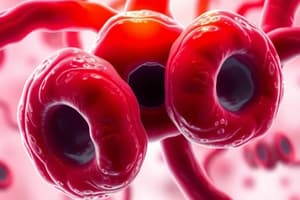
Red Blood Cell (RBC) Variations
- Variations in RBC include size, shape, hemoglobin content, presence of inclusion bodies, and distribution in blood smears.
- Elliptocyte (Ovalocyte): Cigar to egg-shaped, caused by defects in red cell membrane proteins. Associated conditions: Hereditary Elliptocytosis, Megaloblastic anemia, Thalassemia.
Normal Red Blood Cell Characteristics
- Erythrocytes measure 6-8 micrometers in diameter (average 7.5 μm).
- Thickness ranges from 1.5-2.5 micrometers (average 2 μm).
- Shape is round and biconcave, known as discocyte, with salmon pink coloration and central pallor.
Types of Abnormal RBC Morphology
- Codocyte (Target Cell): Bull's-eye appearance, associated with liver disease, hemoglobinopathies, Thalassemia.
- Anisocytosis: Increased variation in RBC size.
- Microcytic RBC: Smaller than normal, linked to iron deficiency anemia and thalassemia.
- Macrocytic RBC: Larger than normal, often due to defects in nuclear maturation, seen in Megaloblastic anemia.
- Echinocyte (Crenated Cell): Regular spicules; changes in osmotic pressure, associated with liver disease.
- Drepanocyte (Sickle Cell): Crescent-shaped; results from hemoglobin S polymerization, associated with hemoglobinopathies.
Specific RBC Shapes
- Spherocyte: Lacks central pallor; caused by spectrin deficiency, associated with hereditary spherocytosis and G6PD deficiency.
- Dacryocyte (Teardrop Cell): Pear-shaped, seen in primary myelofibrosis and thalassemia.
- Stomatocyte (Mouth Cell): Slit-like central pallor, caused by osmotic changes and associated with Rh null, alcoholism.
- Hyperchromasia: Deeply stained RBCs with abnormal thickness, observed in macrocytosis and megaloblastic anemia.
Erythrocyte Inclusion Bodies
- Basophilic Stippling: Aggregated RNA within red cells, visualized by Wright and supravital stains, associated with lead poisoning and severe anemia.
- Cabot Rings: Found in hemolytic and megaloblastic anemia.
- Pappenheimer Bodies: Dark-staining iron particles, aggregates of mitochondria and ribosomes, associated with hemoglobinopathies.
Nucleated RBCs
- Indicate bone marrow stimulation or increased erythropoiesis; associated with hemolytic anemias and myeloproliferative disorders.
Alterations in RBC Distribution
- Rouleaux Formation: Stacking of RBCs.
- Agglutination: Clumping of RBCs.
Parasitic Inclusions in Erythrocytes
- Plasmodium falciparum: Malignant tertian malaria, normal size with Maurer’s dots.
- Plasmodium ovale: Benign tertian malaria, normal to slightly enlarged, with Schuffer’s dots.
- Plasmodium vivax: Benign tertian malaria, enlarged RBCs with Schuffner’s dots.
- Plasmodium malariae: Quartan malaria, normal size with Ziemann’s dots.
Grading of Hypochromia
- 1+: Area of central pallor is ½ diameter of the cell.
- 2+: Central pallor is 2/3 of cell diameter.
- 3+: Central pallor is ¾ of cell diameter.
- 4+: Thin rim of hemoglobin.
Grading of Polychromasia
- 1+: 3% of red cells are polychromatophilic.
- 2+: 5%.
- 3+: 10%.
- 4+: >11%.
Studying That Suits You
Use AI to generate personalized quizzes and flashcards to suit your learning preferences.