Podcast
Questions and Answers
Which type of potentials do photoreceptors, bipolar cells, horizontal cells, and amacrine cells use?
Which type of potentials do photoreceptors, bipolar cells, horizontal cells, and amacrine cells use?
- Graded potentials (correct)
- Electrical conduction
- Action potentials
- Synaptic potentials
What type of conduction do retinal neurons use to conduct their visual signals?
What type of conduction do retinal neurons use to conduct their visual signals?
- Neurotransmitter conduction
- Electrical conduction (correct)
- Ion channel conduction
- Chemical conduction
What is the result of the hyperpolarization (due to the sodium-calcium channels closure) during the activation of photoreceptors by light?
What is the result of the hyperpolarization (due to the sodium-calcium channels closure) during the activation of photoreceptors by light?
- Depolarization of the membrane
- Decreased glutamate release (correct)
- Increased sodium-calcium channel opening
- Release of neurotransmitters
What is the function of glutamate in photoreceptors?
What is the function of glutamate in photoreceptors?
What happens to cGMP in the dark?
What happens to cGMP in the dark?
What is the result of the closure of sodium-calcium channels in photoreceptors' light activation?
What is the result of the closure of sodium-calcium channels in photoreceptors' light activation?
What is the function of transducin in photoreceptors?
What is the function of transducin in photoreceptors?
What type of cells generate action potentials?
What type of cells generate action potentials?
Which of the following is NOT a factor that affects visual acuity?
Which of the following is NOT a factor that affects visual acuity?
What is the purpose of the lens in the eye?
What is the purpose of the lens in the eye?
What is the characteristic of images formed on the retina?
What is the characteristic of images formed on the retina?
What is the purpose of the retina in the process of image formation?
What is the purpose of the retina in the process of image formation?
What is the difference in the way cone cells and rod cells feed signals to ganglion cells?
What is the difference in the way cone cells and rod cells feed signals to ganglion cells?
What is the characteristic of the central retina?
What is the characteristic of the central retina?
What happens to the light rays emanating from the top and bottom of the object as they approach the eye?
What happens to the light rays emanating from the top and bottom of the object as they approach the eye?
What is the function of the optic radiation?
What is the function of the optic radiation?
What is the role of the pretectal nucleus in the pupillary light reflex arc?
What is the role of the pretectal nucleus in the pupillary light reflex arc?
What is the function of the cones and rods in the retina?
What is the function of the cones and rods in the retina?
Choose the correct order of fiber termination for visual reflexes.
Choose the correct order of fiber termination for visual reflexes.
What is the order of light refraction in the eye?
What is the order of light refraction in the eye?
What is the function of the optic nerve?
What is the function of the optic nerve?
Which species have the highest percentage of cranial nerve 2 (optic nerve) decussation?
Which species have the highest percentage of cranial nerve 2 (optic nerve) decussation?
Which of the following is not an example of an effector organ used for the pupillary light reflex?
Which of the following is not an example of an effector organ used for the pupillary light reflex?
Flashcards are hidden until you start studying
Study Notes
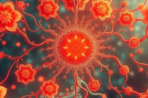
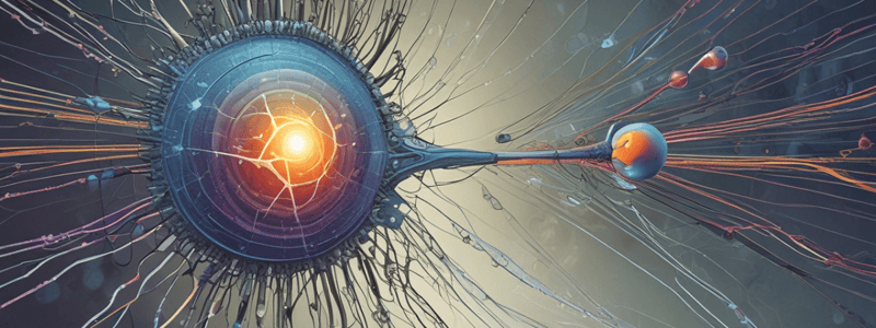
Activation of Photoreceptors by Light
- Photoreceptors, bipolar cells, horizontal cells, and amacrine cells use graded potentials rather than action potentials.
- These cells conduct their visual signals along the cell by direct flow of electric currents (electrical conduction).
- The strength of the hyperpolarizing output signal is directly related to the intensity of the illumination.
Activation of Photoreceptors by Light: Steps
- Step 1: Activation of rhodopsin by light leads to activation of a G-protein, transducin.
- Step 2: The activated transducin splits away and activates PDE6.
- Step 3: The activated PDE6 hydrolyzes cGMP to 5'-GMP (inactive form), causing the sodium-calcium channel to close.
- Step 4: The sodium-calcium channel closure leads to hyperpolarization (membrane potential becomes more negative).
- Step 5: The decrease in glutamate released will excite the bipolar cells.
Photoreceptors in the Dark
- In the dark, cGMP phosphodiesterase is inactive (PDE6).
- cGMP accumulates and binds to ligand-gated sodium-calcium ion channels.
- The flow of cations into the rods keeps the membrane depolarized.
Visual Acuity
- Acuity of visual images reflects the following factors: population of retinal cells, ratio of rods to cone photoreceptor cells, and ratio of photoreceptor cells to ganglion cells.
- Cone cells provide better acuity than rod cells.
- Hundreds of rods feed signals via bipolar cells to a single ganglion cell.
- Only a few cone cells feed signals to a single ganglion cell.
Image Formation: General Info
- Images are formed by the eye, which is optically equivalent to a photographic camera.
- The eye catches the light reflected by objects and guides its passage until the image is formed.
- The lens works by converging the light rays to a certain focal point on the retina.
Image Formation: Light Refraction Steps
- Step 1: Light is refracted off of the interface between the air and the anterior surface of the cornea.
- Step 2: Light is refracted at the interface between the posterior surface of the cornea and the aqueous humor.
- Step 3: Light is refracted at the interface between the aqueous humor and the anterior surface of the lens of the eye.
- Step 4: Light is refracted at the interface between the posterior surface of the lens and the vitreous humor.
Visual Pathway
- The images formed on the retina are transformed by cones and rods into nerve impulses.
- The brain processes and reorients the image.
- Visual signals are carried by optic nerve fibers that form the optic nerve, optic chiasm, and optic tract.
Pupillary Light Reflex (PLR)
- The PLR is a reflex arc composed of receptors for light within the retina, afferent neurons, brainstem centers, and efferent neurons.
- The PLR is involved in pupillary constriction.
Studying That Suits You
Use AI to generate personalized quizzes and flashcards to suit your learning preferences.