Podcast Beta
Questions and Answers
What is the significance of the resonance hybrid in peptide bonds?
Which of the following statements is true about the dihedral angles φ and ψ?
What characterizes the α-helix structure?
Which amino acid is typically considered a helix breaker?
Signup and view all the answers
What does the term 'random coil' refer to in protein structure?
Signup and view all the answers
How do side chains affect the stability of the α-helix?
Signup and view all the answers
What does the tertiary structure of a protein refer to?
Signup and view all the answers
What type of bonds play a significant role in stabilizing the tertiary structure of proteins?
Signup and view all the answers
What is characterized by the arrangement of two or more separate polypeptide chains?
Signup and view all the answers
Which of the following is an example of a fibrous protein?
Signup and view all the answers
How do two α-helices form a coiled coil in α-keratin?
Signup and view all the answers
What strengthens the quaternary structure of α-keratin?
Signup and view all the answers
What is the primary characteristic of fibrous proteins?
Signup and view all the answers
Which process is involved in the biochemical engineering of permanent waving?
Signup and view all the answers
What is the primary role of collagen in connective tissues?
Signup and view all the answers
Which amino acid is most abundant in collagen?
Signup and view all the answers
What structural feature is characteristic of myoglobin?
Signup and view all the answers
What is the function of ascorbic acid in collagen synthesis?
Signup and view all the answers
How many α-helices are present in myoglobin?
Signup and view all the answers
What is the main role of human serum albumin in the blood?
Signup and view all the answers
What percentage of myoglobin's structure is composed of α-helices?
Signup and view all the answers
Which structure is characterized by having a high alpha-helix content?
Signup and view all the answers
What type of bond is responsible for cross-linking collagen fibrils?
Signup and view all the answers
What is the consequence of oxidation of Fe2+ in myoglobin?
Signup and view all the answers
What defines a motif in the context of protein structure?
Signup and view all the answers
How many subunits does hemoglobin contain for its quaternary structure?
Signup and view all the answers
Which protein has the highest percentage of β-conformation as per the provided data?
Signup and view all the answers
What is the term for proteins that assist in the proper folding of other proteins?
Signup and view all the answers
What configuration is formed by the three separate polypeptides in collagen?
Signup and view all the answers
Which of the following statements about protein denaturation is true?
Signup and view all the answers
Study Notes
Peptide Bond Structure
- The peptide bond is a resonance hybrid of two canonical structures.
- Resonance causes the peptide bond to be less reactive compared to esters, rigid and nearly planar (but flexible), and to exhibit a large dipole moment, in the favored trans configuration.
Peptide Plane and Rotations
- Rotation around the peptide bond is not permitted.
- Rotation around bonds connected to the alpha carbon is permitted.
- ϕ (phi): angle around the α-carbon—amide nitrogen bond.
- ψ (psi): angle around the α-carbon—carbonyl carbon bond.
- In a fully extended polypeptide both ϕ and ψ are 180°.
- The polypeptide is made up of a series of planes linked at α carbons.
Distribution of ϕ and ψ Dihedral Angles
- Some ϕ and ψ combinations are unfavorable, due to steric crowding of backbone atoms with other atoms in the backbone or side chains.
- Some ϕ and ψ combinations are more favorable, due to the chance to form favorable H-bonding interactions along the backbone.
- Some ϕ and ψ values are impossible due to steric clash.
- Ramachandran plots help to visualize possible and impossible ϕ and ψ angles.
Secondary Structure
- Refers to a local spatial arrangement of the polypeptide backbone.
- Two regular arrangements are common: α helix and β sheet.
- The α helix is stabilized by hydrogen bonds between nearby residues.
- The β sheet is stabilized by hydrogen bonds between adjacent segments that may not be nearby.
- Irregular arrangement of the polypeptide chain is called the random coil.
- Can be determined experimentally through circular dichroism spectroscopy.
The α Helix
- The helical backbone is held together by hydrogen bonds between the backbone amides of an n and n+4 amino acids (CO group of AA1 with NH group of AA5).
- Right-handed helix with 3.6 residues (5.4 Å) per turn.
- Peptide bonds are aligned roughly parallel with the helical axis.
- Side chains point out and are roughly perpendicular with the helical axis.
- ϕ = -57°
- ψ = -47°
The α Helix (cont.)
- α-helix sequence can be represented through the helix wheel, which provides a top view, indicating connectivity but also the positioning of the AA residues along the helix.
- Amino acids have different propensities to form α-helices, depending on their R group properties.
- Small hydrophobic residues such as Ala and Leu are strong helix formers.
- Pro acts as a helix breaker because the rotation around the N-Ca bond is impossible.
- Gly acts as a helix breaker because the tiny R-group supports other conformations.
- Attractive or repulsive interactions between side chains 3–4 amino acids apart will affect formation → the position of an AA relative to its neighbors is important!
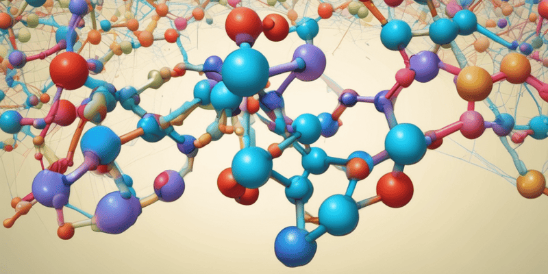
Protein Tertiary Structure
- The overall 3D arrangement of all atoms in a protein is referred as the protein’s tertiary structure.
- Includes long-range aspects of the amino acid sequence (AA that are far apart in polypeptide sequence may interact within the completely folded 3D structure of the protein).
- Interacting segments of polypeptide chains are held in their characteristic tertiary positions by weak interactions and by disulfide covalent bonds.
Protein Quaternary Structure
- Some proteins contain two or more separate polypeptide chains.
- The arrangement of these protein subunits in 3D complexes constitutes quaternary structure.
- Exemplified for fibrous proteins: have structural role in organism, consist usually largely of a single type of secondary structure (e.g. α-keratin, collagen, silk fibroin).
Fibrous Proteins: Structure of α-Keratin
- Evolved for strength; found in hair, wool, nails, claws, quills, horns, hooves, outer layer of skin.
- Two α-helices assemble to form a coiled coil, further assemble into protofilaments, protofibrils.
- Strength enhances by covalent cross-links made out of disulfide bonds that stabilize quaternary structure.
Structure of α-Keratin (cont.)
- Permanent waiving is a biochemical engineering process that involves reducing, moist heat (shaping i.e. straightening or curling), and oxidizing.
Structure of Collagen
- Collagen evolved to provide strength in connective tissue, cartilages, tendons, organic matrix of the bone, cornea.
- Collagen contains a very high proportion of Gly (35%) and Pro (21%); also 4-OH Pro, Ala).
- Left-handed helix, with 3 AA per turn (usually Gly-Pro-4OH-Pro).
- Three separate polypeptides are super-twisted about each other in a unique tertiary and quaternary structure:
Structure of Collagen (cont.)
- 4OH-Pro is biosynthesized from Pro by proline hydroxylase, which requires ascorbic acid (vitamin C).
- Collagen fibrils cross-linked by imine bonds between modified Lys and 5-OH Lys.
Globular Proteins: Structure of Myoglobin
- Myoglobin is a globular protein containing a single polypeptide (153 AAs) and a single iron protoporphirin (heme) group.
- Contains 8 α-helices, interrupted by bends, some of which are β-turns.
- Abundant in muscles, has oxygen transport role through coordination of O2 molecule by Fe2+ (ferrous form).
- Heme is placed in a hydrophobic crevice created by α-helices to prevent oxidation of Fe2+ to Fe3+ (does not bind O2), which occurs in the presence of water.
Approximate Proportion of α Helix and β Conformation in Some Single-Chain Proteins
- Table 4-4 shows the approximate proportion of α helix and β conformation in some single-chain proteins.
Globular Proteins (cont.)
- Human serum albumin is another globular protein.
- It is an important protein in blood plasma; its role is maintaining osmotic pressure, and transporting fatty acids, steroids, and thyroid hormones.
- It has a very high α-helix content.
- Hemoglobin is a globular protein (multimer), whose quaternary structure has 4 almost identical subunits (oligomers), each sub-unit similar to myoglobin.
- It is the main transporter of O2 in blood (red blood cells).
Special Ternary Structures of DNA Binding Proteins
- Helix-Turn-Helix, Zinc Fingers, and Leucine Zipper motifs are supersecondary structures that define recognizable folding patterns involving two or more elements of secondary structure and the connection between them.
- Domain: part of polypeptide chain that is independently stable or could undergo movements as a single entity with respect to the entire protein.
Protein Denaturation and Folding
- Maintaining the protein's native conformation is essential for its function, this is called proteostasis.
- Requires permanent protein synthesis and folding, refolding.
- Folding can occur spontaneously, or can involve specialized proteins called chaperones (e.g. heat shock proteins HSP, chaperonins).
Protein Misfolding and Formation of Amyloid Fibrils
- Misfolding can trigger aggregation, which leads to the formation of fibrils; accumulation of these fibrils can trigger diabetes, Parkinson’s and Alzheimer’s diseases.
Protein Denaturation and Renaturation
- Loss of protein structure results in loss of function; this process is called denaturation of proteins.
- Proteins can be denatured by heat, extreme pH, addition of amphiphilic solvents (such as EtOH, acetone), addition of certain solutes (such as urea and guanidine hydrochloride), and addition of detergents.
- The tertiary structure of proteins is encoded in the AA sequence, which is directly proven by the renaturation of proteins (return to native conformation once denaturant is removed).
Studying That Suits You
Use AI to generate personalized quizzes and flashcards to suit your learning preferences.
Related Documents
Description
Test your knowledge on the structure and properties of peptide bonds. This quiz explores the rigidity of peptide bonds, the allowed rotations around the alpha carbon, and the significance of ϕ and ψ dihedral angles in polypeptide chains. Discover how steric factors and hydrogen bonding influence these angles.