Podcast
Questions and Answers
The nervous tissue is ectodermal in origin.
The nervous tissue is ectodermal in origin.
True (A)
Which of the following is NOT part of the central nervous system?
Which of the following is NOT part of the central nervous system?
- Spinal cord
- Vertebral column
- Brain
- Nerves (correct)
The neuron is the structural and functional unit of the nervous tissue.
The neuron is the structural and functional unit of the nervous tissue.
True (A)
What are the two main parts of a neuron?
What are the two main parts of a neuron?
Dendrites carry impulses away from the cell body.
Dendrites carry impulses away from the cell body.
Nissl's granules are present in the axon.
Nissl's granules are present in the axon.
What is the term for the breakdown of Nissl's granules after injury or exhaustion?
What is the term for the breakdown of Nissl's granules after injury or exhaustion?
Sensory neurons carry impulses towards the CNS.
Sensory neurons carry impulses towards the CNS.
What is the function of interneurons?
What is the function of interneurons?
All nerve fibers have one or two sheaths.
All nerve fibers have one or two sheaths.
The myelin sheath is formed by Schwann cells in both the CNS and PNS.
The myelin sheath is formed by Schwann cells in both the CNS and PNS.
Golgi type I fibers are very short
Golgi type I fibers are very short
Myelinated fibers contain Nissl's granules.
Myelinated fibers contain Nissl's granules.
What type of synapse is most common?
What type of synapse is most common?
A nerve ganglion is a collection of nerve cell bodies inside the CNS.
A nerve ganglion is a collection of nerve cell bodies inside the CNS.
Cranio-spinal ganglia are autonomic.
Cranio-spinal ganglia are autonomic.
Cranio-spinal ganglia have a thin capsule.
Cranio-spinal ganglia have a thin capsule.
The nerve fibers in cranio-spinal ganglia are typically myelinated.
The nerve fibers in cranio-spinal ganglia are typically myelinated.
Synapses are present in cranio-spinal ganglia
Synapses are present in cranio-spinal ganglia
Microglia are ectodermal in origin.
Microglia are ectodermal in origin.
Astrocytes are star-shaped cells with multiple short processes.
Astrocytes are star-shaped cells with multiple short processes.
Oligodendrocytes produce myelin sheaths in the PNS.
Oligodendrocytes produce myelin sheaths in the PNS.
Microglia are derived from blood monocytes.
Microglia are derived from blood monocytes.
Flashcards
Nervous Tissue Origin
Nervous Tissue Origin
Nervous tissue is derived from ectodermal cells.
Central Nervous System (CNS)
Central Nervous System (CNS)
Part of the nervous system comprising the brain and spinal cord.
Peripheral Nervous System (PNS)
Peripheral Nervous System (PNS)
Part of the nervous system outside the brain and spinal cord, including nerves and ganglia.
Neuron
Neuron
Signup and view all the flashcards
Neuron Parts
Neuron Parts
Signup and view all the flashcards
Neuron Cell Body (Perikaryon)
Neuron Cell Body (Perikaryon)
Signup and view all the flashcards
Nissl Bodies
Nissl Bodies
Signup and view all the flashcards
Axon
Axon
Signup and view all the flashcards
Dendrites
Dendrites
Signup and view all the flashcards
Sensory Neuron
Sensory Neuron
Signup and view all the flashcards
Motor Neuron
Motor Neuron
Signup and view all the flashcards
Myelin Sheath
Myelin Sheath
Signup and view all the flashcards
Neuroglia (Glial Cells)
Neuroglia (Glial Cells)
Signup and view all the flashcards
Astrocytes
Astrocytes
Signup and view all the flashcards
Synapse
Synapse
Signup and view all the flashcards
Study Notes
Nervous Tissue Overview
- Nervous tissue is of ectodermal origin
- Anatomically, the nervous system divides into:
- Central nervous system (CNS):
- Brain (inside the skull)
- Spinal cord (inside the vertebral column)
- Peripheral nervous system (PNS):
- Nerves
- Nerve ganglia
- Nerve endings
- Central nervous system (CNS):
- Structurally, nervous tissue consists of two cell types:
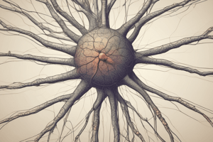
- Nerve cells (neurons):
- The human nervous system has over 100 million neurons
- Neurons are the structural and functional unit
- Functions:
- Reception and transmission of stimuli
- Triggering of cellular activities
- Glial cells (neuroglia):
- Support, protection, and nutrition of neurons in the CNS
- Nerve cells (neurons):
Neuron Structure
- Neurons have two main parts:
- Cell body (perikaryon): Contains the nucleus and surrounding cytoplasm
- Processes:
- Dendrites
- Axon
- Internal structures of the neuron:
- Nucleus: Single, spherical, central, fine dispersed euchromatin, prominent nucleolus
- Cytoplasm: Mildly basophilic, contains cell organelles and inclusions
- Nissl's bodies (granules): Highly developed rough endoplasmic reticulum (rER) with free ribosomes and polyribosomes, important for protein synthesis; not found in the axon
- Golgi complex: Stacks of membranes around the nucleus
- Mitochondria: Present in all parts of the neuron
- Neurofilaments (intermediate filaments): Present in all parts of the neuron, stained with silver
- Microtubules: Present in all parts of the neuron; involved in support, shaping, and impulse transport.
- Lysosomes
- Cell inclusions (e.g., lipid, glycogen, lipofuscin, melanin): Stored food and pigments
Axons and Dendrites
- Axon:
- Arises from the perikaryon in a short pyramidal region called the axon hillock
- Lacking Nissl's granules
- Gives terminal arborizations (branches) in the PNS
- Axolemma is the plasma membrane of the axon and axoplasm is the interior cytoplasm
- Contains mitochondria, microtubules, neurofilaments, and axoplasmic vesicles
- Dendrites:
- Usually multiple and short
- Branch like tree branches, similar cytoplasm to perikaryon
- Lack Golgi complexes but have mitochondria, Nissl bodies, neurofilaments and microtubules
- Carry impulses towards the cell body
Classification of Nerve Cells (Neurons)
-
Based on the number of processes:
- Unipolar (in invertebrates)
- Pseudo-unipolar (spinal ganglion neurons)
- Bipolar (retina and olfactory mucosa neurons)
- Multipolar (most neurons)
-
Based on function:
- Motor neurons (efferent): Carry impulses from CNS to effectors
- Sensory neurons (afferent): Receive sensory stimuli and carry them towards the CNS
- Interneurons: Connect other neurons in the CNS
Nerve Fibers
- Consist of axons enveloped by special sheaths
- Cellular sheath (neurolemmal sheath):
- Formed by Schwann cells in the PNS
- Important for nerve fiber regeneration
- Myelin sheath:
- Formed by Schwann cells in PNS
- Interrupted tube surrounding axons
- Helps in conduction and insulation of nerve impulses
Synapse
- Site of contact between two neurons or neuron and effector cell
- Types of synapses:
- Axodendritic
- Axosomatic
- Axoaxonic
Nerve Ganglia
- Collections of nerve cell bodies outside the CNS
- Ovoid structures surrounded by connective tissue capsule
- Contain neuronal cell bodies and glial cells
Craniospinal Ganglia and Autonomic Ganglia
- Craniospinal ganglia (sensory):
- Unipolar neurons on spinal and cranial nerve roots
- No synapses within the ganglia
- Autonomic ganglia:
- Multipolar neurons
- Impulse transmission from CNS to ganglia in the sympathetic and parasympathetic systems (intramural ganglia)
- Differences between the two types: (summarized)
- Structure: Capsule thickness and shape differences, number and distribution of neurons and nuclei, and presence or absence of satellite cells; nerve fiber myelin features, and synapses.
Neuroglia/Glial Cells
- Types: Astrocytes, Oligodendrocytes, Microglia, Ependymal cells
- Astrocytes:
- Star-shaped cells with multiple processes
- End foot processes at capillaries
- Mildly basophilic cytoplasm with gliosomes (lysosomes)
- Oligodendrocytes:
- Fewer processes than astrocytes
- Form myelin sheaths in the CNS
- Central, oval shaped nucleus
- Microglia:
- Mesodermal origin, monocytes of the blood
- Small and elongated with branching processes
- Basophilic cytoplasm with lysosomes
- Ependymal cells: Ectodermal in origin; form epithelial lining of brain ventricles
Studying That Suits You
Use AI to generate personalized quizzes and flashcards to suit your learning preferences.