Podcast
Questions and Answers
What is the primary artery responsible for supplying blood to the structures in the orbit, including the eyeball?
What is the primary artery responsible for supplying blood to the structures in the orbit, including the eyeball?
- Central retinal artery
- Supraorbital artery
- Ophthalmic artery (correct)
- Inferior palpebral artery
What occurs if the central retinal artery is occluded?
What occurs if the central retinal artery is occluded?
- Improved vision
- Increased intraocular pressure
- Blindness (correct)
- Loss of peripheral vision
Which of the following arteries enters the eyeball by piercing the sclera?
Which of the following arteries enters the eyeball by piercing the sclera?
- Muscular arteries
- Lacrimal artery
- Anterior ciliary artery
- Long and short posterior ciliary arteries (correct)
Which branch of the ophthalmic artery supplies the lacrimal gland and related muscles?
Which branch of the ophthalmic artery supplies the lacrimal gland and related muscles?
What is characteristic of the central artery of the retina in terms of its connections with other arteries?
What is characteristic of the central artery of the retina in terms of its connections with other arteries?
What are the two main components of the neural-sensory layer of the retina?
What are the two main components of the neural-sensory layer of the retina?
Which layer of the retina is primarily responsible for visual resolution and contains only cones?
Which layer of the retina is primarily responsible for visual resolution and contains only cones?
What is the primary purpose of the optic disc?
What is the primary purpose of the optic disc?
Which layer of the retina is directly adjacent to the photoreceptor cells?
Which layer of the retina is directly adjacent to the photoreceptor cells?
What does the term 'pars caeca retinae' refer to?
What does the term 'pars caeca retinae' refer to?
Which artery is the first branch of the ophthalmic artery that supplies the retina?
Which artery is the first branch of the ophthalmic artery that supplies the retina?
What is a defining feature of the central retinal vessels?
What is a defining feature of the central retinal vessels?
What is the role of photoreceptor cells in the retina?
What is the role of photoreceptor cells in the retina?
Which layer of the retina is responsible for the initial processing of visual information?
Which layer of the retina is responsible for the initial processing of visual information?
What structure marks the transition between the optic part of the retina and the nonvisual part?
What structure marks the transition between the optic part of the retina and the nonvisual part?
Which artery supplies blood to the ciliary processes and the iris?
Which artery supplies blood to the ciliary processes and the iris?
Which of the following statements about the retina is incorrect?
Which of the following statements about the retina is incorrect?
What is the correct sequence of the aqueous humor flow in the eye?
What is the correct sequence of the aqueous humor flow in the eye?
Which muscle of the iris is responsible for pupil dilation?
Which muscle of the iris is responsible for pupil dilation?
The optic part of the retina is sensitive to light primarily due to which component?
The optic part of the retina is sensitive to light primarily due to which component?
Where do the efferent impulses responsible for pupil constriction originate?
Where do the efferent impulses responsible for pupil constriction originate?
Study Notes
Vessels
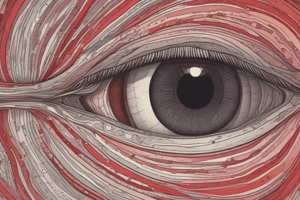
- The ophthalmic artery supplies the orbit, including the eyeball.
- The ophthalmic artery is a branch of the internal carotid artery, originating after the internal carotid artery leaves the cavernous sinus.
- The ophthalmic artery enters the orbit through the optic canal along with the optic nerve.
- The ophthalmic artery runs inferior and lateral to the optic nerve, then crosses superior to the optic nerve.
- The ophthalmic artery proceeds anteriorly on the medial side of the orbit.
Ophthalmic Artery Branches:
- Lacrimal artery: located on the lateral side of the optic nerve, supplying the lacrimal gland, muscles, and anterior ciliary branch.
- Central retinal artery: supplies the center of the optic nerve to the retina. Occlusion of this vessel or its parent artery leads to blindness.
- Long and short posterior ciliary arteries: enter the eyeball posteriorly, piercing the sclera, and supplying structures inside the eyeball. The central artery of the retina is an end artery without effective anastomoses, making occlusion a cause of blindness.
- Muscular arteries:
- Supraorbital artery: originates from the ophthalmic artery, exiting the orbit through the supraorbital foramen with the nerve.
Neural- Sensory layer
- Composed of two layers:
- Outer—PIGMENTED LAYER
- Inner---NERVOUS LAYER
Ora Serrata
- The point where the nervous layer of the retina ends.
- The pigmented layer continues anteriorly, covering the ciliary processes and iris.
- ciliary part
- iridial part
Neural Tunic (Retina)
- The retina from the optic disc to the ora serrata has ten layers containing photoreceptor cells (rods and cones). The layers, from outside to inside (continuous with the optic nerve) are:
- Pigmented epithelium
- Layer of rods and cones
- Outer limiting membrane
- Outer nuclear layer
- Outer plexiform layer
- Inner nuclear layer
- Inner plexiform layer
- Ganglion cell layer
- Optic nerve fiber layer
- Inner limiting membrane
Parts of the Retina
- Optic part: extends from the optic disc to the ora serrata. Contains pigmented and nervous layers.
- Pars caeca retina: also known as the "blind part" of the retina. Located anteriorly from the ora serrata and contains only the pigmented layer.
- Macula lutea: a yellowish, oval area near the center of the posterior part of the retina. It contains the:
- Fovea centralis: a central depression in the macula lutea with high visual resolution. It only contains cones.
- Optic disc:
- Located 3 mm nasal to the macula lutea.
- The point where the optic nerve enters the eyeball. The lamina cribrosa is the point of entry.
- The optic disc is an elevation on the lamina cribrosa.
Optic Disc
- The optic disc is also referred to as the "blind spot" as it is insensitive to light.
- The central artery and vein of the retina are normally pink under ophthalmoscopic examination.
- If the optic nerve atrophies, the color will appear white.
Central Retinal Artery
- This artery runs through the optic nerve fibers.
- It is the first branch of the ophthalmic artery.
- Branches:
- Superior and inferior branches
- Each branch divides into temporal and nasal branches.
- Each quadrant of the retina is supplied by four arteries.
- No anastomoses exist.
Central Retinal Vessels
- These vessels are the only ones in the body that can be inspected directly with the naked eye using an ophthalmoscope.
Iris
- The colored portion of the eye (blue to dark brown).
- Surrounds the pupil.
- It is an adjustable diaphragm.
- The iris has two margins:
- Ciliary
- Pupillary
- The iris is attached to the cornea by pectinate ligaments.
Iris
- It is somewhat suspended in the aqueous humor and divides the area between the cornea and lens into two chambers:
- Anterior chamber
- Posterior chamber
- The cornea and iris meet at the iridocorneal angle.
Aqueous Humor
- Flow: ciliary processes --> posterior chamber --> pupil --> anterior chamber --> sinus venosus sclerae (at the iridocorneal angle) --> anterior ciliary veins --> ophthalmic veins --> cavernous sinus.
Muscles of the Iris
- Sphincter pupillae muscle:
- Smooth muscle responsible for meiosis (pupil constriction).
- Innervated by the parasympathetic system through short ciliary nerves (oculomotor nerve).
- Dilator pupillae muscle:
- Smooth muscle responsible for mydriasis (pupil dilation).
- Innervated by the sympathetic system through the superior cervical sympathetic ganglion, internal carotid artery, long ciliary nerves (nasociliary nerve) (or short ciliary nerves).
Arteries of the Iris
- Long posterior ciliary arteries
- Anterior ciliary arteries
- Arteries of the ciliary process
- Major arterial circle (at the ciliary margin)
- Minor arterial circle (at the pupillary margin)
Inner Layer of the Eyeball (Retina)
- The inner layer of the eyeball is the retina.
- The retina consists of two parts:
- Optic part of the retina: posteriorly and laterally, this part of the retina is sensitive to light.
- Nonvisual part: anteriorly, this part of the retina covers the internal surface of the ciliary body and the iris.
- The junction between the optic and nonvisual parts is an irregular line called the ora serrata.
Optic Part of the Retina
- The optic part of the retina consists of two layers:
- Outer pigmented layer
- Inner neural layer.
- The pigmented layer is firmly attached to the choroid and continues anteriorly, covering the internal surface of the ciliary body and iris.
- The neural layer is only attached to the pigmented layer around the optic nerve and the ora serrata.
Nervous Layer (Retina)
- This is the innermost layer of the eyeball.
Studying That Suits You
Use AI to generate personalized quizzes and flashcards to suit your learning preferences.
Related Documents
Description
This quiz focuses on the anatomy of the ophthalmic artery, including its origin, pathways, and branches. Test your knowledge on the supply of the orbit and the critical functions of its branches, such as the lacrimal artery and central retinal artery.