Podcast
Questions and Answers
The synaptic cleft at the NMJ is approximately 20um wide, allowing direct contact between the nerve and muscle.
The synaptic cleft at the NMJ is approximately 20um wide, allowing direct contact between the nerve and muscle.
False
An alpha motor neuron is also referred to as a lower motor neuron.
An alpha motor neuron is also referred to as a lower motor neuron.
True
Neurotransmitter ACh binds to receptors on the presynaptic terminal of the NMJ.
Neurotransmitter ACh binds to receptors on the presynaptic terminal of the NMJ.
False
The process of neurotransmitter release involves the transfer of signals across the synaptic cleft without physical contact between the nerve and muscle.
The process of neurotransmitter release involves the transfer of signals across the synaptic cleft without physical contact between the nerve and muscle.
Signup and view all the answers
Skeletal muscle contraction involves the process known as cross-bridge formation.
Skeletal muscle contraction involves the process known as cross-bridge formation.
Signup and view all the answers
ACh is produced and stored in synaptic vesicles in the nerve terminal.
ACh is produced and stored in synaptic vesicles in the nerve terminal.
Signup and view all the answers
Type I muscle fibers are commonly referred to as fast twitch fibers.
Type I muscle fibers are commonly referred to as fast twitch fibers.
Signup and view all the answers
Myofibrils are made up of thick filaments known as actin.
Myofibrils are made up of thick filaments known as actin.
Signup and view all the answers
The sarcomere is the basic contractile unit of skeletal muscle and is defined by Z discs.
The sarcomere is the basic contractile unit of skeletal muscle and is defined by Z discs.
Signup and view all the answers
Fast twitch muscle fibers are more fatigue resistant than slow twitch fibers.
Fast twitch muscle fibers are more fatigue resistant than slow twitch fibers.
Signup and view all the answers
Calcium is stored in the sarcoplasmic reticulum of muscle fibers.
Calcium is stored in the sarcoplasmic reticulum of muscle fibers.
Signup and view all the answers
The action potential in skeletal muscle spreads via a system of T-tubules.
The action potential in skeletal muscle spreads via a system of T-tubules.
Signup and view all the answers
Troponin and tropomyosin are additional protein components associated with thick filaments.
Troponin and tropomyosin are additional protein components associated with thick filaments.
Signup and view all the answers
Study Notes
Learning Outcomes
- Define a synapse
- Distinguish between an alpha motor neuron, lower motor neuron, and somatic motor neuron
- Understand the structure of the neuromuscular junction (NMJ) including its components
- Describe the release of neurotransmitters at the NMJ
- Describe the sequence of events following the release of neurotransmitters at the NMJ
- Recall the names of the different muscle fiber components, like myofibrils
- Define and explain the sequence of events in skeletal muscle contraction (cross-bridge formation) and relaxation
Neuromuscular Junction (NMJ)
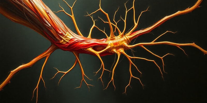
- NMJ is a chemical synapse
- It is where the motor nerve meets the muscle
- The NMJ is about 20um wide with no physical contact between the nerve and the muscle
- The nerve and the muscle are separated by a synaptic cleft
- The neuromuscular cleft is the site of neurotransmitter release
Neurotransmitter Release
- The neurotransmitter acetylcholine (ACh) is released into the synaptic cleft between the nerve and muscle
- ACh is produced and stored in synaptic vesicles in the presynaptic nerve terminal
- Release of ACh is triggered by an action potential in the motor neuron
- ACh binds to receptors on the muscle cell membrane
- Binding of ACh triggers an end-plate potential (EPP) in the muscle cell membrane
Muscle Contraction
- The EPP triggers an action potential in the muscle fiber
- This action potential spreads throughout the muscle fiber through a network of T-tubules
- The action potential causes the release of calcium ions from the sarcoplasmic reticulum (SR)
- Calcium ions bind to troponin, causing a conformational change in tropomyosin
- The conformational change in tropomyosin exposes the binding sites on the actin filaments for myosin heads
Muscle Relaxation
- ACh is broken down by acetylcholinesterase (AChE), which is located in the synaptic cleft.
- The breakdown of ACh stops the signal to the muscle fiber and the process of muscle contraction.
- Calcium ions are pumped back into the SR, which removes them from the sarcomere
- Without calcium ions, the troponin and tropomyosin complex returns to its resting position covering the binding sites
- This stops the interaction between actin and myosin
- Muscle fibers relax.
Muscle Fiber Types
- There are two main types of skeletal muscle fibers: Type I slow twitch (red) and Type II fast twitch (white)
- Type I (red) are fatigue resistant with high myoglobin, mitochondria, and oxidative metabolism
- Type II (white) are fatigue prone, with less myoglobin, mitochondria, and oxidative metabolism, but are more glycolytic
- Oxidative metabolism uses oxygen to produce energy, and glycolytic metabolism doesn't use oxygen
Muscle Structure
- Muscle fibers are made up of myofibrils
- Myofibrils are made up of myofilaments of actin (thin filaments) and myosin (thick filaments)
- Myofibrils have a striated appearance
- The contractile unit of skeletal muscle is the sarcomere
Sarcomere
- Extends from one Z disc to the next
- Contains a variety of different proteins
- Actin: Thin filament
- Myosin: Thick filament
- Titin
- Troponin and Tropomyosin: additional protein components of the actin filament
Muscle Contraction and Relaxation Cycle
- The interaction of actin (thin) and myosin (thick) filaments within the sarcomere is essential for muscle contraction
- Calcium ions play a critical role in regulating the interaction between actin and myosin
Studying That Suits You
Use AI to generate personalized quizzes and flashcards to suit your learning preferences.
Related Documents
Description
This quiz covers key aspects of the neuromuscular junction (NMJ) including the structure, function, and processes involved in muscle contraction. You'll explore components like alpha motor neurons, neurotransmitter release, and the sequence of events that lead to skeletal muscle contraction and relaxation.


