
30 Questions
What is the primary function of mitochondria in a cell?
To extract energy from food substances and store it in ATP
What is the significance of the highly folded inner membrane of mitochondria?
It increases the surface area for ATP production
Which type of cells are likely to have a high abundance of mitochondria?
Skeletal muscle cells
What is the energy stored in ATP molecules?
In the bond between the phosphate group and the rest of the molecule
What happens when a phosphate group is 'snapped off' an ATP molecule?
The bond is broken and energy is released
What is the result of re-attaching a phosphate group to an ADP molecule?
The molecule becomes ATP
What is the main function of skeletal muscle?
To bring about movement
What gives skeletal muscle its striated appearance?
The highly regular arrangement of actin and myosin
What is the function of connective tissue in skeletal muscle?
To group muscle fibers together
Where are the nuclei of skeletal muscle cells located?
On the outer surface of the cell
What surrounds the whole muscle?
The muscle sheath
What type of control is skeletal muscle under?
Voluntary or conscious control
What is the primary structure that attaches skeletal muscle to a bone?
Tendon
What are the two contractile proteins that make up a myofibril?
Actin and myosin
What is the arrangement of muscle cells or fibres?
Long and cylindrical, lying parallel to each other
What is the function of the connective tissue in skeletal muscle?
To provide structure and support to the muscle fibres
Where are the nuclei of skeletal muscle cells located?
On the outer surface of the cell
How are muscle fibres grouped?
Into bundles or fascicles by connective tissue
What is the primary characteristic of skeletal muscle cells or fibres?
They are long and cylindrical.
What is the function of the muscle sheath?
To surround and protect the muscle.
What is the arrangement of myofibrils in skeletal muscle cells?
Highly regular and parallel.
What is the location of the nuclei in skeletal muscle cells?
On the outer surface of the cell.
What is the type of control that skeletal muscle is under?
Voluntary or conscious.
What are the two contractile proteins that make up a myofibril?
Actin and myosin.
What is the characteristic that gives skeletal muscle its striated or striped appearance when viewed under a microscope?
The arrangement of myofibrils
Skeletal muscle is under involuntary control.
False
What is the function of the muscle sheath?
It surrounds the whole muscle and is continuous with the tendons that connect the muscle to a bone.
The muscle fibres are grouped together in bundles or __________ by connective tissue.
fascicles
Match the muscle components with their descriptions:
Myofibrils = Microfilaments that give skeletal muscle its striated appearance Actin = Thin filaments that make up myofibrils Myosin = Thick filaments that make up myofibrils Connective tissue = Holds muscle fibres together
Each muscle fibre has a single nucleus.
False
Study Notes
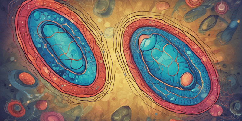
Mitochondria
- Responsible for cellular respiration and energy extraction from food substances
- Site where energy is stored in the form of ATP (Adenosine triphosphate)
- Have a smooth outer membrane and a highly folded inner membrane
- Increased surface area of the inner membrane enables efficient ATP production
- Found in abundance in cells with high energy consumption, such as skeletal muscle
- Cells use ATP stores when energy is required
- Energy is stored in the bond between the phosphate group and the rest of the ATP molecule
- Breaking the bond by 'snapping off' a phosphate group releases energy
- Remaining molecule is then called adenosine diphosphate (ADP) with two phosphate groups
- Re-attaching a phosphate group (during metabolic processes) restores energy storage as ATP
Characteristics of Skeletal Muscle
- Found attached to the skeleton and enables movement
- Under voluntary or conscious control, allowing animals to move their limbs
- Muscle cells or fibers are long and cylindrical and lie parallel to each other
- Each individual muscle fiber is composed of bundles of microfilaments known as myofibrils
Structure of Muscle Fibers
- Myofibrils are made of two contractile proteins: actin (thin filaments) and myosin (thick filaments)
- The regular arrangement of these filaments gives the muscle its striated or striped appearance under a microscope
Muscle Fiber Composition
- Each fiber has several nuclei located on the outer surface of the cell
- Myofibrils push cell structures to the outer margins, resulting in nuclei at the surface
Muscle Organization
- Muscle fibers are grouped together in bundles or fascicles by connective tissue
- Groups of fascicles are held together by connective tissue to form a large muscle
- The entire muscle is surrounded by the muscle sheath, which connects to tendons that attach to bones
Learn about mitochondria, the site of cellular respiration, and how they extract energy from food substances to produce ATP. Explore their structure and function in active cells.
Make Your Own Quizzes and Flashcards
Convert your notes into interactive study material.
Get started for free


