Podcast
Questions and Answers
Blockage in the normal drainage of lymph can result in which of the following conditions?
Blockage in the normal drainage of lymph can result in which of the following conditions?
- Lymphedema (correct)
- Lymphopoiesis
- Leukocytosis
- Lymphoma
Which characteristic distinguishes lymph capillaries from blood capillaries?
Which characteristic distinguishes lymph capillaries from blood capillaries?
- Lining by stratified squamous epithelium
- Blind-end tubes with flattened or irregular shaped lumen (correct)
- Presence of valves to prevent backflow
- Thicker walls with multiple layers of cells
What is the function of mini-valves in lymphatic capillaries?
What is the function of mini-valves in lymphatic capillaries?
- Directing lymph flow away from tissues
- Regulating blood pressure within the capillary
- Preventing the entry of fluids and large solutes
- Permitting entry of fluids, solutes, viruses, bacteria, and cell debris (correct)
The superficial and deep lymph collecting vessels converge to form larger vessels known as:
The superficial and deep lymph collecting vessels converge to form larger vessels known as:
Which of the following is a primary function of the lymphatic system?
Which of the following is a primary function of the lymphatic system?
Which characteristic is associated with innate immune defenses?
Which characteristic is associated with innate immune defenses?
What role do interferons play in chemical defense?
What role do interferons play in chemical defense?
Which process enables phagocytes to leave capillaries and enter infected tissues?
Which process enables phagocytes to leave capillaries and enter infected tissues?
What is the role of immunological surveillance?
What is the role of immunological surveillance?
Which class of lymphocytes is responsible for antibody-mediated immunity?
Which class of lymphocytes is responsible for antibody-mediated immunity?
How do antibodies contribute to opsonization?
How do antibodies contribute to opsonization?
Which characteristic describes adaptive immune defenses?
Which characteristic describes adaptive immune defenses?
What is the role of Class I MHC proteins?
What is the role of Class I MHC proteins?
What is the function of helper T cells?
What is the function of helper T cells?
Which type of antibody is primarily found in body secretions such as mucus, tears, and saliva?
Which type of antibody is primarily found in body secretions such as mucus, tears, and saliva?
Flashcards
Immune System
Immune System
Complex collection of cells and organs that destroys or neutralizes pathogens.
Lymphatic System
Lymphatic System
System of vessels, cells, and organs carrying excess fluids to bloodstream and filtering pathogens.
Lymph
Lymph
Tissue fluid that enters lymphatic vessels.
Lacteals
Lacteals
Signup and view all the flashcards
Chyle
Chyle
Signup and view all the flashcards
Barrier Defenses
Barrier Defenses
Signup and view all the flashcards
Innate Immune Response
Innate Immune Response
Signup and view all the flashcards
Adaptive Immune Response
Adaptive Immune Response
Signup and view all the flashcards
Lymphocytes
Lymphocytes
Signup and view all the flashcards
Lymphopoiesis
Lymphopoiesis
Signup and view all the flashcards
Primary Lymphoid Organs
Primary Lymphoid Organs
Signup and view all the flashcards
Secondary Lymphoid Organs
Secondary Lymphoid Organs
Signup and view all the flashcards
Buboes
Buboes
Signup and view all the flashcards
Lymph Nodules
Lymph Nodules
Signup and view all the flashcards
Innate Defenses
Innate Defenses
Signup and view all the flashcards
Study Notes
- The immune system consists of cells and organs that destroy pathogens
- The lymphatic system consists of vessels, cells, and organs and moves excess fluid to the bloodstream and filters pathogens.
Functional Anatomy of the Lymphatic and Immune Systems
- The lymphatic system drains excess interstitial fluid from tissues and transports it to the bloodstream
- Lymph is tissue fluid that has entered lymphatic vessels
- Lymphedema is produced by blockage of lymph drainage
- Phagocytic cells and lymphocytes housed by this system clean tissue fluid before it enters circulation
- Lacteals are specialized lymph vessels that absorb fats from the intestine
- Chyle is fatty lymph
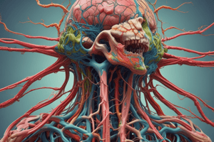
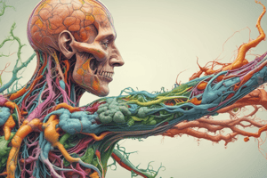
Structure of the Lymphatic System
- The lymphatic system includes vessels, cells, tissues, and organs
- Lymph vessels carry lymph from peripheral tissues towards the venous system
- Lymphatic capillaries start the lymphatic network and merge into progressively larger vessels for circulation
- Lymph capillaries are present in almost every tissue and organ, thought to be as abundant as blood capillaries
- Lymph capillaries are absent in areas lacking a blood supply, like the cornea, the central nervous system, and bone marrow.
- Lymph capillaries differ from blood capillaries:
- They are blind-ended, have larger diameters, and possess thinner, more permeable walls.
- They also typically have flattened or irregular lumens.
- Lymphatic capillaries have simple squamous epithelium linings and incomplete or absent basal lamina
- Endothelial cells overlap to form mini-valves, permitting fluids and solutes (proteins) entry while preventing their return
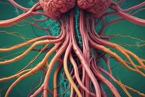
- Lymph flows from the capillaries into larger lymph collecting vessels equipped with valves, directing lymph toward the body's trunk
- Lymph trunks form from the convergence of superficial and deep lymph collecting vessels and are named after the body areas they drain:
- Jugular trunks: neck, drain the head
- Subclavian trunks: shoulders, drain the arms
- Bronchomediastinal trunks: chest, drain the thoracic cavity and lungs
- Lumbar trunks: lower back, drain the pelvis and lower limbs
- Intestinal trunk: abdomen, drains the walls of the digestive organs
- Blockage of lumbar or subclavian trunks by filarial worms (Wuchereria bancrofti) results in severe lymphedema (Elephantiasis)
- Lymph ducts form from the merger of lymph trunks, creating the right lymphatic duct and thoracic duct
- The right lymphatic duct forms from the merger of the right jugular, subclavian, and bronchomediastinal trunks
- Drains the right side of the head, arm, shoulder and thoracic cavity and empties into the right subclavian vein
- The thoracic duct ascends along the left side of the vertebral column and collects lymph from the left bronchomediastinal, subclavian, and jugular trunks
- Cisterna chyli is the enlarged sac-like base of the thoracic duct that receives lymph from the lumbar and intestinal trunks
- Drains the left side of the head, arm, and shoulder, as well as the thoracic cavity, abdomen, pelvic region, and both legs, and empties into the left subclavian vein
Organization of Immune Function
-
Barrier defenses: These are the skin and mucous membranes, provide instantaneous action against pathogenic invasion
-
Innate immune response: This is a fast, non-specific defense with specialized cells and soluble factors
-
Adaptive immune response: This response is slower, specific, and effective, uses leukocytes (primarily lymphocytes)
-
Erythropoiesis normally occurs in red bone marrow in adults
-
Lymphocyte production (lymphopoiesis) involves red bone marrow, the thymus, and peripheral lymphoid tissues
-
Functional classes of WBCs:
- Phagocytic cells: ingest destroying pathogens
- Lymphocytes: adaptively coordinate immunity
- Granular WBCs: mediate immune responses against parasites and intracellular pathogens such as viruses
-
Lymphocytes include B cells, T cells, Plasma cells, and Natural Killer cells.
- Lymphocytes account for 20-30% of circulating leukocytes:
- Most reside within lymph organs like the lymph nodes, spleen, tonsils, and thymus
- Lymphocytes account for 20-30% of circulating leukocytes:
Lymphocyte Classes
- B cells account for 10-15% of lymphocytes for antibody-mediated (humoral) immunity:
- Antibodies circulate to attack foreign antigens, B cells differentiate into plasma cells when stimulated
- Plasma cells produce and secrete antibodies, contain cytoplasm packed with protein-synthesizing machinery (rough endoplasmic reticulum)
- T cells constitute ~80% lymphocytes and provide cell-mediated immunity
- Natural Killer (NK) cells constitute 5-10% of lymphocytes for non-specific defenses, attacking foreign cells, virus-infected cells, and cancer cells:
- Immunological surveillance represents continuous monitoring of peripheral tissues
Primary Lymphoid Organs and Lymphocyte Development
- Red bone marrow: Blood cells develop in the yolk sac during embryo. Function is taken over by the spleen, lymph nodes, then the liver until bone marrow takes over:
- Thymocytes: immature T cells leave bone marrow and mature in thymus
- Thymus contains hormones called thymosins important for functional T cell development and normal body defense maintenance
- The thymus grows during childhood when infection increases. By puberty, the thymus declines gradually in size during involution
- By age 50, the thymus may be <12g correlated with infection/disease-susceptibility
- The thymus capsule divides into left/right lobes divided into lobules averaging 2 mm in diameter
- Each lobule contains a dark outer cortex and lighter central medulla, with medulla containing thymic corpuscles not present in the cortex
Secondary Lymphoid Organs
- Naïve lymphocyte: One that that enters a secondary lymphoid organ is fully functional immunologically, but has yet to encounter an antigen
- Lymph nodes: small lymphoid organs 1-25mm in diameter. Large collections reside in cervical axillary, and inguinal regions
- 99% of antigens removed from lymph as lymph flows, stimulating immune response
- Swollen lymph nodes are called buboes
- Lymph flow in lymph node:
- Afferent lymphatic vessels carry lymph into node via the capsule side opposite the hilum where blood vessels and nerves enter/leave
- Afferent vessels deliver lymph to subcapsular space which contains meshwork of reticular fibers macrophages, dendritic cells, playing roll in immune response
- Lymph flows to the outer cortex containing B cells within germinal centers
- Lymph flows through the deep cortex containing T cells - Lymph continues into medullary sinuses at core of the lymph node with B and plasma cells
- Efferent lymphatic vessels drain "cleaned" lymph out of node from hilum
- Spleen: largest lymphoid organ filters blood by removing abnormal RBCs, store iron and activate immune response via B and T activation in bloodstream
- The spleen lies along stomach's lateral border attached by the gastrosplenic ligament and the diaphragmatic surface is smooth
- The spleen is surrounded by fragile capsule containing collagen and elastin fibers making it easy to rupture, leading to splenectomies
- Splenic blood vessels and lymphatic vessels communicate with spleen via medial (visceral) surface at hilum which also contains shallow depressions (gastric/renal area)
- Trabeculae are fibrous partitions which radiate fromhilum and blood vessels travel within them
- Red pulp: The spleen contains large quantities of red blood cells.
- White pulp: The spleen resembles lymphoid nodules and contains lymphocytes and give phagocytes opportunity to engulf infected or damaged cells
Lymph Nodules
- These are areas of densely-packed lymphatic tissue that contain reticular connective tissue with larger numbers of elastin and reticular fibers without a fibrous capsule
- Tonsils: large lymphoid nodules in pharynx walls
- Palatine tonsils: posterior, inferior margin of oral cavity
- Pharyngeal tonsil (adenoid): lies in posterior superior nasopharynx wall
- Lingual tonsils: lie deep in mucous epithelium at tongue base
- Tubal tonsils: found in base of pharyngotympanic tubes
- When infected, our tonsils go unnoticed unless swollen known as tonsillitis
- Mucosa-associated lymphoid tissue (MALT) protects epithelia of the digestive, respiratory, urinary & reproductive tracts from pathogens/toxins
- Appendicitis: inflammation of lymphoid tissue from appendix
- Aggregated lymphoid nodules (Peyer's patches): MALT type clustered deep in distal small intestine
- Bronchus-associated lymphoid tissue (BALT): lymphoid follicular structures
Characteristics of the innate body defenses
- Innate defenses are present and functioning at birth
- Innate defenses are non-specific, responding the same way regardless of threat
- Innate defenses tend to be more localized to attack the infection at entry
- Innate defenses have no memory for improved response upon repeated exposures
Innate Immunity
- Includes physical barriers, cellular defenses via phagocytosis & NK cells, & chemical defenses (complement inflammatory chemicals interferon and pyrogens)
Physical Barriers
- These keep organisms and materials outside the body
- Skin: the integumentary system provides major physical barrier to the external environment
- Epidermis: the skin is composed of stratified squamous epithelium with keratinized cells and a network of desmosomes that lock adjacent cells together
- Hairs provide minimal protection by providing mechanical abrasion protection (especially on the scalp) and prevent hazardous material from insect contact.
- Epidermal surfaces flush surfaces with secretions from sweat glands and contain bactericidal chemicals such as defensins destructive enzymes called Lysosomes, and antibodies
- Epidermal surfaces receive lubricating secretions from sebaceous glands creating an arid hostile environment for microorganisms
- Mucous membranes: epithelial linings of digestive, respiratory, urinary, & reproductive tracts provide barrier
- Mucus bathes membranes trapping organisms and debris, preventing entry
- Mucous membranes secrete chemicals (acids, lysozymes, and defensins) to reduce microorganism growth
- Mucous membrane cells connected by tight junctions and supported by thick fibrous basal lamina
- Mucous membranes possess cilia that create outward movement to transport microorganisms up/out
- Mucous membranes possess MALT
Cellular Defenses
- If a hazardous organism gains entry into the body, cells attack the infection at the site of entry
- Phagocytic cells: must make it past physical barriers to engulf pathogens/debris
Phagocytes
- All phagocytes can leave capillaries through squeezing between endothelial cells during diapedesis or emigration
- Phagocytes are attracted to chemicals through positive chemotaxis released by infection
- Phagocytosis begins with adherence where receptors attach on the plasma membrane binding onto surface
- After attachment, phagocyte will destroy and/or promote destruction via T/B cell activation
Different types of phagocytic cells:
- Neutrophils: abundant leukocytes mobilize quickly towards sites of injury phagocytizing debris/bacteria
- Eosinophils: less abundant than neutrophils; have limited abilities to phagocytize until pathogens are coated with antibodies
- Monocytes: give rise to fixed and macrophages
- Free macrophages: travel throughout body, arrive at the site of injury by traveling through adjacent tissues - Fixed macrophages: permanent residents of CT
NK(natural killer) cells
- These cells detect and destroy abnormal body cells/virus-infected cells unless cancer cells develop. NK cells constantly monitor and recognize bacteria foreign-infected cells
- Immunological surveillance: constant monitoring
- Step one: cells with unusual components recognized as abnormal NK cells adhere to cell
- Step two: large number of perforins, a series of proteins which travel towards cell surface
- Step one perforins at cell surface exocytosis and diffuses in narrow gap separating NK cells and target
- Step one perforins create holes/pores in plasma membrane, the target loses internal environment
Chemical Defenses
- Chemical substances that destroy organism label it invading, prevent reproduction /stimulate immune system response
- Interferons: cytokine secreted by lymphocytes macrophages, tissues infected by viruses
- If a virus is present in the body interferons protect healthy cells causing them to bind on membrane via secondary messenger
- Alpha interferons: product of viral resistance to infection and attracts NK cells
- Beta interferons: secreted by fibroblast for slowing inflammation
- Gamma interferons: secreted by T and NK cells for stimulate macrophage activity
Complement
- Made of 11 circulating protein, assist/complement antibodies on pathogens
- Classical pathway: most rapid and effective occurs when the complement attaches to antibody molecules already bound to their specific antigen
- Alternative pathway: important on defense against bacteria parasites and Factor B, factor D, and properdin
Inflammatory Chemicals
- These control localized tissue to avoid spread
- Step one: tissue damage caused by infections or temperature induce
- Step two: cause cytokines potassium and pyrogens
- Step three: Basophilis that release histamine(vascular permeability) and heparin(vasodilation)
- Vasodilation: increases blood flow,
- Permeability: increases causing exudate,
- Fibers threads: occlude lymphs,
- Leukocytosis– increased cell production
- Pyrogens: fever that elevates body temperature to accelerates metabolic activities and promotes activity
- Fever increase metabolism inhibited by an increase in temperature
Adaptive Immune Response
- These involve lymphocytes that are not present at birth and must be developed, be sensitive for an infection, and have memory
- Versatile: Clone adapt to multiply based on clone and produce different responses on site
- tolerance: Immune system that tolerates normal tissues by targeting abnormal
Cell Mediated Immunity
- It’s based on action of T-lymphocytes in bone marrow and matures in the Thymus gland and cannot recognized antigen
- Instead APC-Antigen presenting cells and the Major histocompatibility complexes or MHC proteins
- Two classes MHC I: on class 1 MHC proteins attach to infection and identity the attacker Class II MHC: alerts body that infection had found
Cell Stimulation and Clonal expansion
- when macrophages patrolling body tissues see foreign organisms, they engulf them via phagocytosis. The Antigen Class II MCH protein:
- When viruses are identified a cell is infected to trigger T- Cells, Tc cells and cell memory
Four types of T-Cell
- Helper T CD-4 cells- include all other for stimulus ( B-Cells and phagocytosis)
- Cytotoxic T-Cells includes killer cells to infect various cells
- Suppressor T reduce down the action when infection stopped
- Memory T-Cells- Remains in circulation until another exposure occurs
Antibody Mediated Immunity
- Function of B- Cells produce and matured in red bone marrow called Humoral Immunity
- B-lymphocytes, reside in spleen lymph nodes circulate infection/antigen Clonal expansion with an antibody structure: 4 polypeptide chains (heavy/ light).
Variable structure
- Constant: amino same sequence variable regions
- the amino acid binds during a particular way
Antigen-Antibody
- Bind Antigen not all components only expose sites: antigen binding sites. a) Antigen Determinates a specific binding region b) Complete- is an antigen with 2 Ag binding sites c) Partial antigen is a hapten ( does ordinarly cause activation Five different classes and immunoglobins structure.
Type of Antibodies
- IgA-dimer and attacks surface of pathogens
- IgD: monomer and where it binds antigen
- IgE attaches for inflammation
- IgG :Resistance from bacterial toxicities, viruses and passes to placenta
- IgM- antibody (anti class) during anti B,
Sensitization
- prepares activation with Antigen. a clonal B -Secretes antibodies from free antigens Memory-Quickly exposure future infections.
Antigen response
- Active: Body produce response and gives long term stability
Innate
- Response causes an automatic acquired for a period
Antibody Mechanism
- Activate: on binding to antigen exposing complement
- Phagocytosis: Antigens collect ( eosinphilitis that Phagocitize to destroy plasma membrane.
- Neutralization- Both viruses and bacterial must bound to plasma membrane to protect bodies bodies
- Inflammation Antibodies causes release of stimulation release to the chemicals found on skin
Immunity system Disorders
- Immune Deficiency- immune system cells
- Severe combined immunodeficiency syndromes with deficitis
- Acquired immune deficiency syndromes : HIV
- Allergies Severe response is to much
Hypersensitivity
- Type I Begins within seconds for an anaphalectic. -Type II and III occur 1-3 hours Type IV: skin irritation and last days
Autoimmune disease
- occurs B when calls attack Antibodies and are numerous:
- Sclerosis Multiple: nervous
- Rheumatoid: joints
- Syematic : attacks organs
Transplantation
- Occurs when tissues are incompatible attack
- Autografts
- Isographs
- Allografts
-
- Xeno-grapphts
Studying That Suits You
Use AI to generate personalized quizzes and flashcards to suit your learning preferences.