Podcast
Questions and Answers
Which of the following is NOT a primary function of the hypodermis?
Which of the following is NOT a primary function of the hypodermis?
- Insulation
- Shock absorption
- Vitamin D production (correct)
- Energy storage
Which layer of the hair structure is the outermost?
Which layer of the hair structure is the outermost?
- Cuticle (correct)
- Medulla
- Cortex
- Hair Bulb
The epidermis is composed of adipose tissue.
The epidermis is composed of adipose tissue.
False (B)
Arrector pili muscles are responsible for sebum production.
Arrector pili muscles are responsible for sebum production.
What is the primary function of the root plexus?
What is the primary function of the root plexus?
What two main cell types are found in the epidermis?
What two main cell types are found in the epidermis?
Which layer of the dermis is superficial?
Which layer of the dermis is superficial?
The fine, soft hair that covers a fetus in the womb is called ______.
The fine, soft hair that covers a fetus in the womb is called ______.
The skin constitutes approximately ______ % of body weight.
The skin constitutes approximately ______ % of body weight.
The epidermis is described as avascular. What does avascular mean?
The epidermis is described as avascular. What does avascular mean?
Match the following hair types with their descriptions:
Match the following hair types with their descriptions:
The reticular dermis makes up approximately 50% of the dermis.
The reticular dermis makes up approximately 50% of the dermis.
Match the skin layer with its description:
Match the skin layer with its description:
Which type of hair is most associated with secondary sexual characteristics?
Which type of hair is most associated with secondary sexual characteristics?
What is the primary tissue type found in the reticular dermis?
What is the primary tissue type found in the reticular dermis?
If epidermal regeneration takes 28–30 days in adults, approximately how many times will someone's epidermis regenerate if they live to be 90 years old?
If epidermal regeneration takes 28–30 days in adults, approximately how many times will someone's epidermis regenerate if they live to be 90 years old?
__________ lines are deep creases found in areas such as palms, wrists, and soles.
__________ lines are deep creases found in areas such as palms, wrists, and soles.
Sudoriferous glands secrete sebum.
Sudoriferous glands secrete sebum.
Match the following pigments with their contribution to skin color:
Match the following pigments with their contribution to skin color:
What is the term for hereditary, hormone-driven hair thinning?
What is the term for hereditary, hormone-driven hair thinning?
Insanely Difficult: Tactile epithelial cells primarily function in melanin production, influencing skin pigmentation and protection against UV radiation.
Insanely Difficult: Tactile epithelial cells primarily function in melanin production, influencing skin pigmentation and protection against UV radiation.
Which of the following is NOT a function of the hypodermis?
Which of the following is NOT a function of the hypodermis?
What is the primary component of sweat secreted by sudoriferous glands?
What is the primary component of sweat secreted by sudoriferous glands?
Sebaceous glands secrete sebum via ______ secretion.
Sebaceous glands secrete sebum via ______ secretion.
What type of tissue primarily composes the hypodermis?
What type of tissue primarily composes the hypodermis?
Hair is found everywhere on the body, including the palms and soles of the feet.
Hair is found everywhere on the body, including the palms and soles of the feet.
What is the function of epidermal ridges?
What is the function of epidermal ridges?
What is the primary component of nails?
What is the primary component of nails?
Which type of gland is primarily responsible for thermoregulation through the production of odorless, watery sweat?
Which type of gland is primarily responsible for thermoregulation through the production of odorless, watery sweat?
Apocrine glands directly open onto the skin surface like eccrine glands.
Apocrine glands directly open onto the skin surface like eccrine glands.
What specific virus is responsible for causing warts on the skin?
What specific virus is responsible for causing warts on the skin?
A first-degree burn affects only the upper ______ of the skin.
A first-degree burn affects only the upper ______ of the skin.
Assuming complete destruction of cutaneous receptors, a massive third-degree burn exposes the patient to what most lethal threat?
Assuming complete destruction of cutaneous receptors, a massive third-degree burn exposes the patient to what most lethal threat?
Which layer of the epidermis is responsible for providing mechanical stress resistance and also houses dendritic cells?
Which layer of the epidermis is responsible for providing mechanical stress resistance and also houses dendritic cells?
Melanocytes are exclusively found in the stratum basale.
Melanocytes are exclusively found in the stratum basale.
What is the primary function of keratinocytes in the epidermis?
What is the primary function of keratinocytes in the epidermis?
The stratum __________ is only found in thick skin, such as the palms and soles of the feet.
The stratum __________ is only found in thick skin, such as the palms and soles of the feet.
Match the epidermal cell types with their primary function:
Match the epidermal cell types with their primary function:
Which of the following is NOT a function of keratinocytes?
Which of the following is NOT a function of keratinocytes?
Hyperpigmentation is caused by underactive melanocytes.
Hyperpigmentation is caused by underactive melanocytes.
What is the mnemonic to remember the layers of the epidermis from superficial to deep?
What is the mnemonic to remember the layers of the epidermis from superficial to deep?
If a patient is diagnosed with vitiligo, which type of melanocyte disorder are they experiencing?
If a patient is diagnosed with vitiligo, which type of melanocyte disorder are they experiencing?
Lamellar granules within the stratum granulosum contain a waterproofing __________.
Lamellar granules within the stratum granulosum contain a waterproofing __________.
Flashcards
Integumentary System
Integumentary System
The organ system that includes skin, hair, nails, and glands.
Skin
Skin
The body's largest organ, making up ~7% of body weight.
Epidermis
Epidermis
The outermost layer of the skin, acting as a protective barrier.
Dermis
Dermis
Signup and view all the flashcards
Functions of the Skin
Functions of the Skin
Signup and view all the flashcards
Hypodermis
Hypodermis
Signup and view all the flashcards
Keratinocytes
Keratinocytes
Signup and view all the flashcards
Melanocytes
Melanocytes
Signup and view all the flashcards
Medulla
Medulla
Signup and view all the flashcards
Cortex
Cortex
Signup and view all the flashcards
Cuticle
Cuticle
Signup and view all the flashcards
Hair follicle
Hair follicle
Signup and view all the flashcards
Arrector Pili Muscle
Arrector Pili Muscle
Signup and view all the flashcards
Vellus hair
Vellus hair
Signup and view all the flashcards
Terminal hair
Terminal hair
Signup and view all the flashcards
Androgenetic Alopecia
Androgenetic Alopecia
Signup and view all the flashcards
Sebaceous Glands
Sebaceous Glands
Signup and view all the flashcards
Sudoriferous Glands
Sudoriferous Glands
Signup and view all the flashcards
Dendritic Cells
Dendritic Cells
Signup and view all the flashcards
Hyperpigmentation
Hyperpigmentation
Signup and view all the flashcards
Hypopigmentation
Hypopigmentation
Signup and view all the flashcards
Stratum Corneum
Stratum Corneum
Signup and view all the flashcards
Stratum Lucidum
Stratum Lucidum
Signup and view all the flashcards
Stratum Granulosum
Stratum Granulosum
Signup and view all the flashcards
Stratum Spinosum
Stratum Spinosum
Signup and view all the flashcards
Stratum Basale
Stratum Basale
Signup and view all the flashcards
Eccrine Gland
Eccrine Gland
Signup and view all the flashcards
Apocrine Gland
Apocrine Gland
Signup and view all the flashcards
First-Degree Burn
First-Degree Burn
Signup and view all the flashcards
Basal Cell Carcinoma
Basal Cell Carcinoma
Signup and view all the flashcards
Acne Vulgaris
Acne Vulgaris
Signup and view all the flashcards
Papillary Dermis
Papillary Dermis
Signup and view all the flashcards
Reticular Dermis
Reticular Dermis
Signup and view all the flashcards
Epidermal Ridges
Epidermal Ridges
Signup and view all the flashcards
Cleavage Lines
Cleavage Lines
Signup and view all the flashcards
Flexure Lines
Flexure Lines
Signup and view all the flashcards
Melanin
Melanin
Signup and view all the flashcards
Carotene
Carotene
Signup and view all the flashcards
Nails
Nails
Signup and view all the flashcards
Hair Structure
Hair Structure
Signup and view all the flashcards
Study Notes
Integumentary System Overview
- The integumentary system is the body's largest organ, accounting for approximately 7% of body weight.
- It's roughly 1.5 to 4.4 millimeters thick.
- It has two main layers: the epidermis and the dermis.
- The subcutaneous tissue (hypodermis) lies below the dermis.
- The integumentary system includes skin, hair, nails, and glands.
- Its functions include protection, temperature regulation, excretion, vitamin D production, and sensory reception.
- The skin's structure and conditions change throughout life.
Learning Objectives
- Students will identify major components of the integumentary system and their functions.
- Students will identify major structures of the skin, glands, and their functions.
- Students will describe the role of dermal circulation in sensing touch, pressure, and pain.
- Students will explain hair and nail structure, functions, and growth processes.
- Students will discuss the specialization of mammary glands as integumentary glands.
Skin and Subcutaneous Tissue
- The skin is the body's largest organ.
- The subcutaneous layer (hypodermis) lies below the dermis.
- The hypodermis comprises areolar and adipose tissues.
- Functions of the hypodermis include insulation, shock absorption, energy storage, and anchoring the skin.
Skin
- The epidermis is the outermost layer of the skin.
- The dermis is the layer beneath the epidermis.
- The subcutaneous tissue (hypodermis) lies below the dermis.
Epidermis
- It's the outermost layer of the skin.
- It's a protective barrier between the body and the external environment.
- The epidermal layers include stratum corneum, stratum lucidum (in thick skin only), stratum granulosum, stratum spinosum, and stratum basale (stratum germinativum).
Epidermal Layers
- Stratum corneum: outermost, horny layer of dead keratinocytes.
- Stratum lucidum: clear layer, in thick skin only, comprised of flat, dead keratinocytes.
- Stratum granulosum: few layers of keratinocytes; keratinocytes above lack nutrient access.
- Stratum spinosum: spiny appearance; contains intermediate filaments for strength and flexibility.
- Stratum basale: deepest layer, actively dividing cells attach to dermis, contains tactile epithelial cells and melanocytes; actively dividing cells.
Main Cell Types of the Epidermis
- Keratinocytes: most abundant epidermal cells, create keratin for strength and water resistance; produce antibiotics and enzymes.
- Melanocytes: produce melanin, protect from UV damage, determine skin, hair, and eye color; found in the stratum basale.
- Tactile epithelial cells (Merkel cells): mechanoreceptors for light touch and pressure, located in the basal layer
- Dendritic cells (Langerhans cells): part of the immune system, monitor and process pathogens, protect against infections and skin diseases; found in stratum spinosum
Keratinocytes
- Compose the majority of the epidermis.
- Produce keratin, a fibrous protein important for skin strength and water resistance.
Melanocytes
- Found in the stratum basale
- Produce melanin, which protects against UV damage and determines skin, hair, and eye color.
Tactile Epithelial Cells
- Mechanoreceptors for light touch and pressure.
- Located in the epidermis (basal layer)
Dendritic Cells
- Part of the immune system, crucial for monitoring and processing pathogens.
- Found in the stratum spinosum, protecting against infections and skin diseases.
Melanocyte Disorders
- Hyperpigmentation is a result of overactive melanocytes (e.g., sunspots, melasma).
- Hypopigmentation occurs due to underactive melanocytes (e.g., vitiligo).
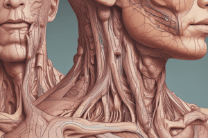
Dermis
- The dermis is a strong, flexible connective tissue layer beneath the epidermis, rich in blood vessels and nerves.
- It comprises the papillary and reticular dermis.
- Papillary dermis: superficial layer.
- Reticular dermis: deeper layer.
Papillary Dermis
- Includes dermal papillae, increasing surface area for gas, nutrient, and waste exchange.
- Nourishes epidermis via capillaries.
- Contains receptors for light touch and vibration.
- Regulates body temperature.
Reticular Dermis
- Dense irregular connective tissue.
- Provides most of the skin's strength.
- Contains collagen and elastic fibers, responsible for the skin's flexibility.
- Contains vascular plexuses for nutrient delivery and temperature regulation (dermal plexus, subpapillary plexus).
- Contains nerve receptors for pressure and pain.
- Composed of cleavage lines.
Cleavage Lines
- Cleavage lines represent separation between underlying collagen fibers in the reticular dermis.
- Surgical incisions parallel to cleavage lines heal better than those made across them.
Flexure Lines
- Flexure lines are deep creases, crucial for flexibility. They occur in areas with significant dermal attachments to underlying structures like palms, wrists, soles, fingers, and toes.
Hypodermis/Subcutaneous Tissue
- Located beneath the skin.
- Made of areolar and adipose tissues.
- Anchors skin, providing insulation, storing energy, and offering cushioning and protection.
- Distribution varies between sexes.
Skin Color
- Melanin: main pigment produced from tyrosine.
- Carotene: yellow-orange pigment from foods.
- Hemoglobin: oxygenated blood gives a rosy undertone to light-skinned individuals.
Appendages of the Skin
- Nails
- Hair
Nails
- Scale-like epidermal modifications made of hard keratin.
- Free edge extends past fingertips.
- Nail plate covering the nail bed.
- Root at the base of the nail.
- Surrounded by nail folds (skin).
- Eponychium (cuticle): skin around the nail.
Hair
- Flexible strand of dead, keratinized cells.
- Found everywhere except palms and soles.
- Embedded within follicles.
- Shaft projects above the skin's surface.
- Made of tough, durable hard keratin.
Hair Structure
- Medulla (central core)
- Cortex (surrounds medulla)
- Cuticle (outermost layer)
Additional Hair Structures
- Hair follicles: structures in the skin where hair grows.
- Hair bulb: base of hair, contains the matrix (involved in hair growth)
- Root plexus: sensory nerve network around hair bulb.
Arrector Pili Muscle
- Tiny smooth muscle attached to hair follicles.
- Causes hair to stand erect when contracting (goosebumps).
- Plays a role in thermoregulation and protection.
Hair Types and Growth
- Vellus hair: fine, short, lightly pigmented hair found on most parts of the body; present in children and adults.
- Terminal hair: coarse, thick, pigmented hair; develops during puberty, notable on the scalp, eyebrows, eyelashes, armpits, and groin.
- Lanugo: fine, soft, unpigmented hair covering the fetus in the womb; typically sheds before or shortly after birth.
Hair Thinning and Baldness
- Androgenetic alopecia: (hormone-driven) thinning at the crown and hairline (men) or overall thinning (women).
- Alopecia areata: autoimmune disorder with patchy hair loss.
- Telogen effluvium: temporary thinning due to stress, illness, or hormonal changes.
- Age-related thinning: gradual thinning with age, influenced by hormones and genetics.
Sebaceous Glands
- Secrete sebum (an oily substance).
- Found everywhere except palms and soles.
- Primarily associated with hair follicles.
Sebum Function
- Lubrication.
- Barrier function.
- Antimicrobial protection.
- Hair conditioning.
Sudoriferous (Sweat) Glands
- Widely distributed on the body.
- Sweat is a blood filtrate (mostly water with salts and metabolic wastes).
- Two types: eccrine glands, and apocrine glands.
Eccrine Glands
- Most abundant type.
- Located in palms, soles, and forehead
- Produce odourless, watery sweat.
- Open directly onto skin surface, contributing significantly to thermoregulation and skin hydration.
Apocrine Glands
- Located in axillary, anal, and genital regions.
- Open into hair follicles.
- Activated during puberty.
- Produce thicker, milky sweat with a musky odor.
Modified Apocrine Glands
- Ceruminous glands: produce earwax.
- Mammary glands: produce milk, hormonally activated.
- Ciliary glands: lubricate eyelashes.
Integumentary System Conditions
- Burns (first, second, third degree)
- First-degree: affects the epidermis.
- Second-degree: damages the epidermis and upper dermis; often results in blisters.
- Third-degree: destroys all skin layers; can appear white, red, or blackened.
- Skin Cancer:
- Basal cell carcinoma least malignant and most common.
- Squamous cell carcinoma is from keratinocytes of stratum spinosum.
- Melanoma is caused by melanocytes and is the most dangerous type.
Infections
- Acne vulgaris: inflammation of sebaceous glands caused by bacteria and clogged follicles.
- Warts: caused by human papillomavirus (HPV) direct contact or contaminated surfaces.
- Ringworm: is a fungal infection producing a ring-shaped rash through direct skin-to-skin contact.
Aging Skin
- Skin thins and loses elasticity.
- Skin inflammations and rashes become more common.
- The skin becomes more susceptible to infections.
Summary
- The integumentary system acts as a protective barrier against external harm and regulates body temperature.
- It includes skin, hair, nails, and glands.
- The skin is a vital barrier and sensory interface. Aging leads to skin changes and less efficient wound healing.
Studying That Suits You
Use AI to generate personalized quizzes and flashcards to suit your learning preferences.
Related Documents
Description
Test your knowledge of the integumentary system with this quiz. Questions cover skin layers, hair structure, and the functions of different components like the epidermis, dermis, and hypodermis. The quiz also assesses understanding of cell types and tissue composition.