Podcast
Questions and Answers
Which part forms the anterior continuation of the ribs and provides flexible attachment at the articulation with the sternum?
Which part forms the anterior continuation of the ribs and provides flexible attachment at the articulation with the sternum?
What forms the osteocartilaginous thoracic cage?
What forms the osteocartilaginous thoracic cage?
Which muscle separates the ribs and their cartilages in the intercostal spaces?
Which muscle separates the ribs and their cartilages in the intercostal spaces?
Which part of the thoracic wall occupies the intercostal spaces and contains muscles and vessels?
Which part of the thoracic wall occupies the intercostal spaces and contains muscles and vessels?
Signup and view all the answers
What is the function of the typical intercostal nerves?
What is the function of the typical intercostal nerves?
Signup and view all the answers
Study Notes
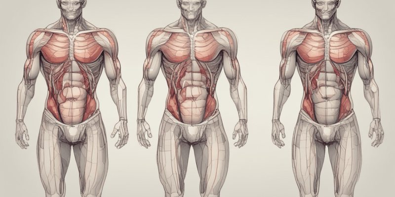
Thoracic Cage Structure
- The costal cartilages form the anterior continuation of the ribs and provide flexible attachment at the articulation with the sternum.
Thoracic Cage Composition
- The osteocartilaginous thoracic cage is formed by the ribs and their cartilages.
Intercostal Space Contents
- The intercostal muscles separate the ribs and their cartilages in the intercostal spaces.
- The intercostal spaces contain muscles and vessels, which are part of the thoracic wall.
Intercostal Nerve Function
- The typical intercostal nerves transmit sensory information from the skin and motor signals to the intercostal muscles.
Studying That Suits You
Use AI to generate personalized quizzes and flashcards to suit your learning preferences.
Description
Test your knowledge of the gross anatomy of the thoracic wall with this quiz. Explore topics such as the skeleton, joints, movements, fascia, and more. Perfect for medical students and professionals seeking to deepen their understanding of thoracic anatomy.