Podcast
Questions and Answers
What is the primary function of the sensory neurons in the nervous system?
What is the primary function of the sensory neurons in the nervous system?
- To activate muscles and glands
- To provide structural support within the nervous system
- To collect sensory input from stimuli (correct)
- To process and interpret sensory information
Which of the following structures is part of the central nervous system?
Which of the following structures is part of the central nervous system?
- Brain (correct)
- Spinal nerves
- Ganglia
- Cranial nerves
What type of matter in the nervous system contains long processes of neurons?
What type of matter in the nervous system contains long processes of neurons?
- Plexus
- Grey matter
- White matter (correct)
- Neuroglia
Which of the following correctly describes synapses?
Which of the following correctly describes synapses?
What is the term for a collection of cell bodies of neurons located outside the central nervous system?
What is the term for a collection of cell bodies of neurons located outside the central nervous system?
Identify the correct sequence of the coverings of the brain and spinal cord from outermost to innermost.
Identify the correct sequence of the coverings of the brain and spinal cord from outermost to innermost.
Which part of the brain is responsible for processing information regarding fine motor skills?
Which part of the brain is responsible for processing information regarding fine motor skills?
What is the main function of cerebrospinal fluid in the central nervous system?
What is the main function of cerebrospinal fluid in the central nervous system?
What is the function of the dorsal root of a spinal nerve?
What is the function of the dorsal root of a spinal nerve?
Which cranial nerve is responsible for vision?
Which cranial nerve is responsible for vision?
Which part of the spinal cord gives rise to the sympathetic division of the autonomic nervous system?
Which part of the spinal cord gives rise to the sympathetic division of the autonomic nervous system?
What characterizes the parasympathetic division of the autonomic nervous system?
What characterizes the parasympathetic division of the autonomic nervous system?
What is a dermatome?
What is a dermatome?
Which of the following statements about the autonomic nervous system is false?
Which of the following statements about the autonomic nervous system is false?
Which cranial nerve is primarily involved in the control of facial expressions?
Which cranial nerve is primarily involved in the control of facial expressions?
What is the axon of the 1st (preganglionic) neuron in the autonomic nervous system responsible for?
What is the axon of the 1st (preganglionic) neuron in the autonomic nervous system responsible for?
Which of the following is a function of the nervous system?
Which of the following is a function of the nervous system?
What type of neuron classification is based on location?
What type of neuron classification is based on location?
Which of the following layers of meninges is the outermost layer?
Which of the following layers of meninges is the outermost layer?
Which part of the brain is NOT included in the major regions of the brain?
Which part of the brain is NOT included in the major regions of the brain?
What does myelination in nervous tissue refer to?
What does myelination in nervous tissue refer to?
What is the origin of the sympathetic division of the autonomic nervous system?
What is the origin of the sympathetic division of the autonomic nervous system?
Which of the following describes the primary function of the parasympathetic division of the autonomic nervous system?
Which of the following describes the primary function of the parasympathetic division of the autonomic nervous system?
What is a dermatome in the context of the nervous system?
What is a dermatome in the context of the nervous system?
Which cranial nerve is not part of the autonomic systems?
Which cranial nerve is not part of the autonomic systems?
Which statement accurately describes the dorsal root of a spinal nerve?
Which statement accurately describes the dorsal root of a spinal nerve?
What is the anatomical and functional unit of the kidney?
What is the anatomical and functional unit of the kidney?
Which part of the urinary bladder is known as the neck?
Which part of the urinary bladder is known as the neck?
How long is the male urethra approximately?
How long is the male urethra approximately?
What is the primary function of the ureters in the urinary system?
What is the primary function of the ureters in the urinary system?
How many nephrons are typically found in each kidney?
How many nephrons are typically found in each kidney?
Which part of the nephron is responsible for reabsorbing nutrients and water?
Which part of the nephron is responsible for reabsorbing nutrients and water?
What is the shape of the urinary bladder when it is empty?
What is the shape of the urinary bladder when it is empty?
What feature distinguishes the male urethra from the female urethra?
What feature distinguishes the male urethra from the female urethra?
Which anatomical feature of the kidneys is associated with the entry and exit points for blood vessels and ureters?
Which anatomical feature of the kidneys is associated with the entry and exit points for blood vessels and ureters?
What is the approximate length of the ureters?
What is the approximate length of the ureters?
Study Notes
Basic Functions of the Nervous System
- Sensation: Monitors internal and external changes (stimuli) using receptors.
- Integration: Processes and interprets sensory information for a proper response.
- Reaction: Activates muscles or glands through neurotransmitters.
Structural Organization of the Nervous System
- Central Nervous System (CNS): Comprises the brain and spinal cord.
- Peripheral Nervous System (PNS): Includes 12 pairs of cranial nerves and 31 pairs of spinal nerves.
Nervous Tissue Composition
- Grey Matter: Contains neuron cell bodies, short processes, neuroglia, and blood vessels.
- White Matter: Composed of long processes of neurons.
Neurons
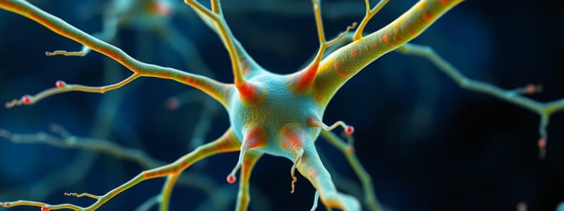
- Definition: Basic units of the nervous system responsible for transmitting impulses.
- Classification: Based on structure, function, and location.
Synapses
- Definition: Junctions where impulses are transmitted between neurons.
- Types: Various classifications exist depending on the functional roles.
Supporting Cells of the Nervous System
- Provide structural and functional support to neurons.
Myelination
- Process of forming a myelin sheath around nerve fibers, enhancing signal transmission speed.
Formation of the Brain and Spinal Cord
- The brain is divided into four main regions:
- Cerebrum: Largest part, consisting of two hemispheres.
- Diencephalon: Contains thalamus and hypothalamus.
- Cerebellum: Involved in coordination and balance.
- Brainstem: Consists of midbrain and pons.
- Spinal Cord:
- Length: 42-45 cm, starts from medulla oblongata to conus medullaris at L2 vertebra and gives rise to 31 pairs of spinal nerves.
Protection of the CNS
- Bones: Skull and vertebral column offer structural protection.
- Meninges: Composed of three layers (dura mater, arachnoid mater, pia mater).
- Cerebrospinal Fluid (CSF): Cushions and protects the brain and spinal cord.
Cranial Nerves
- 12 pairs; key examples include:
- Olfactory Nerve (1st): Smell
- Optic Nerve (2nd): Vision
- Vagus Nerve (10th): Regulation of internal organs
Spinal Nerves
- 31 pairs classified as:
- 8 cervical
- 12 thoracic
- 5 lumbar
- 5 sacral
- 1 coccygeal
- Each spinal nerve has dorsal (sensory) and ventral (motor) roots.
Dermatomes
- Each spinal nerve supplies a specific segment of skin, known as a dermatome.
Autonomic Nervous System (ANS)
- Definition: Regulates involuntary body functions.
- Components: Supplies cardiac and smooth muscle as well as internal organs.
- Neurons: Comprises preganglionic and postganglionic neurons; axons connect CNS to organs.
Divisions of the ANS
- Sympathetic Division: Originates from T1-L2 segments (thoracolumbar), mobilizes energy; activates during stress ("fight or flight").
- Parasympathetic Division: Originates from cranial and sacral regions (craniosacral), conserves energy, promotes maintenance ("rest and digest").
Components of the Urinary System
- Includes kidneys, ureters, urinary bladder, and urethra.
- Two kidneys located on either side of the vertebral column.
Anatomical Structures of Kidneys
- Quantity: Two kidneys, right and left.
- Shape: Bean-shaped.
- Location: Below the ribs, adjacent to the vertebral column.
- Size: Approximately 1 x 2 x 4 inches.
- Structural Features:
- Upper end associated with supra-renal glands.
- Lower end with two distinct borders: outer (convex) and inner (concave with hilum).
- Two surfaces: anterior and posterior.
Nephron Structure
- Quantity: About one million nephrons per kidney.
- Components: Consists of glomerulus and tubule.
- Tubule Differentiation:
- Proximal convoluted tubule
- Loop of Henle
- Distal convoluted tubule
- Collecting duct
Ureters
- Consist of two fibromuscular tubes.
- Length: Approximately 25 cm each.
- Location: Upper part in the abdomen, lower part in the pelvis.
- Function: Transport urine from kidneys to urinary bladder.
Urinary Bladder
- Definition: Hollow muscular organ.
- Location: Situated in the pelvic cavity, behind the symphysis pubis.
- Function: Stores urine.
- Shape: Pyramidal.
- Key Features:
- Apex attached to umbilicus via median umbilical ligament.
- Base oriented posteriorly with inner surface referred to as trigone; receives ureter openings.
- Contains three surfaces and an inferior angle known as the neck, which is surrounded by the internal urethral sphincter (involuntary).
Urethra
Male Urethra
- Length: About 8 inches (20 cm).
- Origin: Starts at neck of the urinary bladder.
- Parts:
- Prostatic: 1.5 inches long.
- Membranous: 0.5 inch long, encased by external sphincter.
- Penile (spongy): 6 inches long.
- Termination: Tip of the glans penis.
Female Urethra
- Length: Around 1.5 inches (4 cm).
- Origin: Begins at the neck of the urinary bladder.
- Termination: Opens into the vestibule in front of the vaginal orifice.
Studying That Suits You
Use AI to generate personalized quizzes and flashcards to suit your learning preferences.
Description
This quiz explores the basic functions of the nervous system, including sensation, integration, and reaction. It also delves into the structural organization of the CNS and PNS, the composition of nervous tissue, and the role of neurons and synapses. Test your understanding of these critical concepts related to nervous system anatomy and function.


