Podcast
Questions and Answers
What is the required posture of the patient when taking a sesamoids tangential view?
What is the required posture of the patient when taking a sesamoids tangential view?
- Patient lying prone or seated (correct)
- Patient standing upright
- Patient lying supine on the table
- Patient in a lateral recumbent position
Which of the following correctly describes the positioning of the foot for a sesamoids tangential view?
Which of the following correctly describes the positioning of the foot for a sesamoids tangential view?
- Foot positioned with the 1st toe extended and ball of foot parallel to the image plate
- Foot positioned resting on the edge of the image plate
- Foot positioned with the 1st toe flexed and ball of foot perpendicular to the image plate (correct)
- Foot positioned with toes parallel to the table edge
For the foot dorso-plantar view, what is the correct angle of the x-ray tube?
For the foot dorso-plantar view, what is the correct angle of the x-ray tube?
- Vertical angle of 0 degrees
- 5 degrees towards the heel
- 10 degrees towards the heel (correct)
- 15 degrees away from the heel
What is the central ray location for a dorso-plantar foot view?
What is the central ray location for a dorso-plantar foot view?
What is a requirement for collimation in a sesamoids tangential view?
What is a requirement for collimation in a sesamoids tangential view?
What should be included in the patient preparation notes prior to taking these foot views?
What should be included in the patient preparation notes prior to taking these foot views?
Which anatomical feature should be visualized in profile without superimposition for the sesamoids tangential view?
Which anatomical feature should be visualized in profile without superimposition for the sesamoids tangential view?
What is the specified mAs range for a sesamoids tangential view?
What is the specified mAs range for a sesamoids tangential view?
What is primarily demonstrated in the imaging of the distal femur and proximal tibia?
What is primarily demonstrated in the imaging of the distal femur and proximal tibia?
Which of the following best describes the appropriate identification of the image?
Which of the following best describes the appropriate identification of the image?
In which scenario would the imaging technique described typically be requested?
In which scenario would the imaging technique described typically be requested?
What does the term 'Horizontal Beam Lateral' indicate in radiographic positioning?
What does the term 'Horizontal Beam Lateral' indicate in radiographic positioning?
Which anatomical structures are primarily visualized in this imaging technique?
Which anatomical structures are primarily visualized in this imaging technique?
What is the appropriate tube angle for a lateral knee view?
What is the appropriate tube angle for a lateral knee view?
Which anatomical structures should be included in the distal femur radiograph?
Which anatomical structures should be included in the distal femur radiograph?
How far should the central ray be positioned for a lateral knee view?
How far should the central ray be positioned for a lateral knee view?
What is the minimum distance that should be maintained from the x-ray tube for a lateral knee projection?
What is the minimum distance that should be maintained from the x-ray tube for a lateral knee projection?
What is the correct positioning for an intercondylar view of the knee?
What is the correct positioning for an intercondylar view of the knee?
What is an important aspect to achieve proper collimation for knee radiographs?
What is an important aspect to achieve proper collimation for knee radiographs?
Which of the following best describes the alignment criteria for a lateral knee view?
Which of the following best describes the alignment criteria for a lateral knee view?
What might affect the density and contrast of a knee radiograph?
What might affect the density and contrast of a knee radiograph?
What is the appropriate kVp range for the Lateral Lower Leg view?
What is the appropriate kVp range for the Lateral Lower Leg view?
Which of the following best describes the posture for the Lateral Lower Leg view?
Which of the following best describes the posture for the Lateral Lower Leg view?
During the AP Knee view, what is the positioning of the knee?
During the AP Knee view, what is the positioning of the knee?
What is the correct central ray positioning for the AP Knee view?
What is the correct central ray positioning for the AP Knee view?
What is required for collimation in both the Lateral Lower Leg and AP Knee views?
What is required for collimation in both the Lateral Lower Leg and AP Knee views?
Which of the following is NOT a criterion for the Lateral Lower Leg view?
Which of the following is NOT a criterion for the Lateral Lower Leg view?
What is the required tube angle for the Lateral Lower Leg view?
What is the required tube angle for the Lateral Lower Leg view?
Which of the following best represents the density criteria for the AP Knee view?
Which of the following best represents the density criteria for the AP Knee view?
What is the primary anatomical focus when performing the intercondylar fossa open view?
What is the primary anatomical focus when performing the intercondylar fossa open view?
What is the optimal tube angle when capturing a skyline view of the patella?
What is the optimal tube angle when capturing a skyline view of the patella?
Which of the following criteria is essential for achieving a good skyline view of the patella?
Which of the following criteria is essential for achieving a good skyline view of the patella?
What is the required postural position for a patient during a PA Rosenberg view?
What is the required postural position for a patient during a PA Rosenberg view?
What mAs range is typically utilized for the patella skyline view?
What mAs range is typically utilized for the patella skyline view?
Which aspect is NOT a requirement in the collimation criteria for the patella skyline view?
Which aspect is NOT a requirement in the collimation criteria for the patella skyline view?
In terms of patient preparation for the skyline view, what is emphasized?
In terms of patient preparation for the skyline view, what is emphasized?
What is the optimal distance for taking a patella skyline view?
What is the optimal distance for taking a patella skyline view?
Flashcards are hidden until you start studying
Study Notes
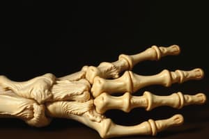
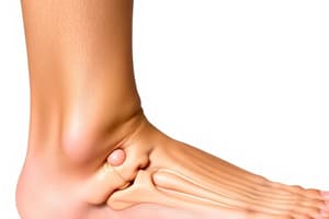
Sesamoids Tangential View
- Position: Patient lying prone or seated with foot positioned with 1st toe flexed and ball of foot perpendicular to image plate. Alternatively, patient can remain seated with foot flexed and tape tensioning toes back.
- Tube Angle: Straight
- Central Ray: Centered on second metatarsal
- Distance: 100-110cm
- Collimation: Include metatarsal head and sesamoids
- kVp: 50-55 kVp
- mAs: 2-4 mAs
- Grid: No
- Patient Prep: Remove artifacts in field of view (shoes, socks)
Sesamoids Tangential View Criteria
- Collimation: Include all required anatomy including skin edges
- Alignment: Joint spaces between sesamoids and 1st metatarsal are open
- Anatomy: Both sesamoids seen in profile without superimposition
- Density: Cortical outline and bony trabecular pattern adequately demonstrated
- Contrast: Soft tissue and bony interfaces visualized
Foot Dorso-Plantar View
- Position: Patient seated on table with foot positioned flat on image plate
- Tube Angle: 10 degrees towards heel
- Central Ray: Centered on base of third metatarsal
- Distance: 100-110cm
- Collimation: Include anatomy of interest
- kVp: 50-55 kVp
- mAs: 3-4 mAs
- Grid: No
- Patient Prep: Remove artifacts in field of view (shoes, socks)
Foot Dorso-Plantar View Criteria
- Collimation: Include all required anatomy including skin edges
- Alignment: No rotation of foot, long axis of image plate aligned with long axis of foot
- Anatomy: Toes to tarsals included, slight overlap of 2nd-5th metatarsal bases
- Density: Cortical outline and bony trabecular pattern adequately demonstrated
- Contrast: Soft tissue and bony interfaces visualized
Lateral Lower Leg View
- Position: Patient lying on side with affected leg outstretched, foot fully flexed, knee slightly bent, true lateral, diagonal image plate to include both joints
- Tube Angle: Straight
- Central Ray: Centered mid shaft tib/fib
- Distance: 100-110cm
- Collimation: Include anatomy of interest
- kVp: 50-60 kVp
- mAs: 3-5 mAs
- Grid: No
- Patient Prep: Remove artifacts in field of view (shoes, clothing)
Lateral Lower Leg Criteria
- Collimation: Include all required anatomy including skin edges
- Alignment: True lateral knee and ankle, slight superimposition of proximal and distal tib/fib
- Anatomy: Knee joint to ankle joint included on one or more images
- Density: Cortical outline and bony trabecular pattern adequately demonstrated
- Contrast: Soft tissue and bony interfaces visualized
AP Knee View
- Position: Patient seated on table with leg extended, knee extended without rotation
- Tube Angle: Straight tube (may use small cephalic angle if patient has big thighs)
- Central Ray: Centered to apex of patella
- Distance: 100-110cm
- Collimation: Include anatomy of interest
- kVp: 60-70 kVp
- mAs: 7-10 mAs
- Grid: No
- Patient Prep: Remove artifacts in field of view (clothing)
AP Knee Criteria
- Collimation: Include all required anatomy including skin edges
- Aignment: No rotation of femoral condyles, symmetry of tibial plateau and centring of tibia intercondylar eminence, some overlap of fibia head on tibia is normal
- Anatomy: Distal femur and proximal tib/fib included, patella superimposed over femur
- Density: Cortical outline and bony trabecular pattern adequately demonstrated
- Contrast: Soft tissue and bony interfaces visualized
Lateral Knee View
- Position: Patient lying on side with knee flexed 30 degrees, unaffected leg positioned in front to assist with adequate rotation and stabilization, knee flexed to engage patella tendon, true lateral, patella in profile
- Tube Angle: 5-7 degrees cephalic angle
- Central Ray: Center on medial aspect of knee joint, 2.5cm distal to medial epicondyle of femur
- Distance: 100-110cm
- Collimation: Include anatomy of interest
- kVp: 60-70 kVp
- mAs: 7-10 mAs
- Grid: No
- Patient Prep: Remove artifacts in field of view (clothing)
Lateral Knee Criteria
- Collimation: Include all required anatomy including skin edges
- Alignment: Both condyles of femur superimposed, patello-femoral joint space open, tibial plateau profiled
- Anatomy: Distal femur, patella and proximal tibia/fibula included
- Density: Cortical outline and bony trabecular pattern adequately demonstrated
- Contrast: Soft tissue and bony interfaces visualized
Intercondylar View
- Position: PA, patient kneeling on stool or AP, patient seated on table with knee bent, AP Seated is more common, knee bent 60 degrees
- Tube Angle: Tube angled to be parallel to tibial plateau
- Central Ray: Cephalic angle to match tibial angle
- Distance: 100-110cm
- Collimation: Include anatomy of interest
- kVp: 60-70 kVp
- mAs: 7-10 mAs
- Grid: No
- Patient Prep: Remove artifacts in field of view (clothing)
Intercondylar Criteria
- Collimation: Include all required anatomy including skin edges
- Alignment: Femoral condyles symmetrical, intercondylar fossa open
- Anatomy: Distal femur and proximal tib/fib included
- Density: Cortical outline and bony trabecular pattern adequately demonstrated
- Contrast: Soft tissue and bony interfaces visualized
Patella Skyline View
- Position: Patient seated on table with knee bent at 30 degrees, skyline is typically an infero-superior axial projection of patella, ideally sitting to ensure quads are unsupported to allow true patella position to be visualized
- Tube Angle: Angle tube to be parallel with patella body
- Central Ray: Centered apex of patella, in plane with patello-femoral space
- Distance: 100-110cm
- Collimation: Include anatomy of interest
- kVp: 60-70 kVp
- mAs: 7-10 mAs
- Grid: No
- Patient Prep: Remove artifacts in field of view (splints, clothing)
Patella Skyline Criteria
- Collimation: Include all required anatomy including skin edges
- Alignment: Superimposition of inferior and superior borders of patella
- Anatomy: Patella profiled axially, patello-femoral joint space open
- Density: Cortical outline and bony trabecular pattern adequately demonstrated
- Contrast: Soft tissue and bony interfaces visualized
PA Rosenberg View
- Position: Patient standing facing upright bucky with knees bent 45 degrees so patellae are in contact with the bucky, tibia and femurs are similar angle from the bucky, don't stand patient too far from the bucky or tibial angle will be too great, often performed bilaterally
- Note: This functional view demonstrates instability on flexion of knees, also useful to visualise a knee prosthesis to check for loosening
- Anatomy: Distal femur, proximal tibia and fibula in lateral profile
- Density: Cortical outline and bony trabecular pattern adequately demonstrated
- Contrast: Soft tissue and bony interfaces visualized
- Markers: Label as “Horizontal Beam Lateral”
- Note: Typically requested in trauma and post operative settings (e.g. after Total Knee Replacement surgery in Recovery room)
Studying That Suits You
Use AI to generate personalized quizzes and flashcards to suit your learning preferences.


