30 Questions
The right atrium sends blood through the Bicuspid valve into the Right ventricle.
False
After passing through the Pulmonary valve, blood enters the Pulmonary trunk.
True
Blood flows from the left ventricle through the Tricuspid valve.
False
The Aorta sends nonoxygenated blood out to the body.
False
Blood flows from the pulmonary veins into the left atrium.
True
Blood sent through the Pulmonary trunk goes directly to the body to gain oxygen.
False
The pressure gradient from high-pressure point to low-pressure point is necessary for fluid flow.
True
Resistance in blood vessels is not affected by factors like blood vessel length, diameter, and viscosity.
False
The AV valves are responsible for producing the 'Lubb' heart sound (S1).
True
During isovolumetric contraction, although ventricles contract, they do not eject blood.
True
The 'Dubb' heart sound (S2) is produced by the closure of the AV valves.
False
During ventricular ejection, blood flows backward into the aorta and pulmonary trunk.
False
The pericardium is a protective tissue surrounding the heart.
True
The pulmonary circuit carries oxygenated blood from the lungs to the heart.
False
The epicardium is another name for the innermost layer of the heart wall.
False
The myocardium is mainly composed of adipose tissue.
False
The endocardium consists of simple squamous epithelium and areolar connective tissue.
True
The apex and base of the heart are represented by an upside-down triangle.
False
The heart has two atria and two ventricles.
True
The coronary sulcus encircles the heart and separates the ventricles from the atria.
False
Semilunar valves prevent the backflow of blood in the heart.
True
The tricuspid valve is also known as the mitral valve and is found on the right side of the heart.
False
Chordae tendineae prevent inversion of the AV valves in the heart.
True
Blood enters the right atrium through the superior vena cava, inferior vena cava, and pulmonary vein.
False
The left coronary artery branches into the anterior interventricular branch and the circumflex branch.
True
The right coronary artery gives rise to the right marginal branch and the posterior interventricular branch.
False
The great cardiac vein is responsible for venous drainage of the heart.
True
The coronary sinus is not involved in the venous drainage of the heart.
False
Intercalated discs in cardiac muscles contain gap junctions that facilitate electrical conduction.
True
The vagus nerve is parasympathetic and speeds up heart rate.
False
Study Notes
Heart Structure
- The heart has two atria and two ventricles.
- The coronary sulcus encircles the heart and separates the ventricles from the atria.
- The pericardium is a protective tissue surrounding the heart.
- The epicardium is another name for the innermost layer of the heart wall.
- The myocardium is not mainly composed of adipose tissue.
- The endocardium consists of simple squamous epithelium and areolar connective tissue.
Blood Flow
- The right atrium sends blood through the Tricuspid valve into the Right ventricle.
- Blood flows from the right ventricle through the Pulmonary valve into the Pulmonary trunk.
- Blood flows from the left ventricle through the Bicuspid valve.
- Blood flows from the pulmonary veins into the left atrium.
- The Aorta sends oxygenated blood out to the body.
Valves
- Semilunar valves prevent the backflow of blood in the heart.
- The Tricuspid valve is not also known as the mitral valve and is found on the right side of the heart.
- Chordae tendineae prevent inversion of the AV valves in the heart.
- The AV valves are responsible for producing the 'Lubb' heart sound (S1).
- The closure of the AV valves produces the 'Dubb' heart sound (S2).
Heart Function
- The pressure gradient from high-pressure point to low-pressure point is necessary for fluid flow.
- Resistance in blood vessels is affected by factors like blood vessel length, diameter, and viscosity.
- During isovolumetric contraction, ventricles contract, but do not eject blood.
- During ventricular ejection, blood flows forward into the aorta and pulmonary trunk.
Blood Vessels and Nerve
- The pulmonary circuit carries deoxygenated blood from the heart to the lungs.
- The left coronary artery branches into the anterior interventricular branch and the circumflex branch.
- The right coronary artery gives rise to the right marginal branch and the posterior interventricular branch.
- The great cardiac vein is responsible for venous drainage of the heart.
- The coronary sinus is involved in the venous drainage of the heart.
- The vagus nerve is parasympathetic and slows down heart rate.
- Intercalated discs in cardiac muscles contain gap junctions that facilitate electrical conduction.
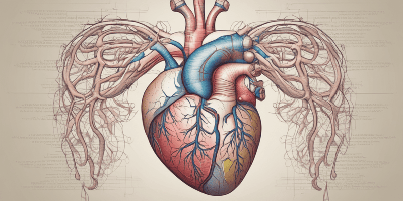
Test your knowledge on how blood flows through the heart, including the role of the right atrium, tricuspid valve, right ventricle, and pulmonary valve. Image source: Saladin, 2018.
Make Your Own Quizzes and Flashcards
Convert your notes into interactive study material.
Get started for free