Podcast
Questions and Answers
What is the branch of science that studies the normal structure of the human body, its organs, the location of these organs, and the relationship between them?
What is the branch of science that studies the normal structure of the human body, its organs, the location of these organs, and the relationship between them?
Anatomy
Which statement about anatomy terminology is correct?
Which statement about anatomy terminology is correct?
- Medical terms have not evolved over time.
- Learning anatomical terminology is fundamental for various health professionals. (correct)
- Anatomy language consists of approximately 10,000 words in the health field.
- Anatomy only uses English terminology.
______ is considered the 'Father of Anatomy' because of his significant contributions to the field.
______ is considered the 'Father of Anatomy' because of his significant contributions to the field.
Vesalius
Match the anatomists with their contributions to anatomy:
Match the anatomists with their contributions to anatomy:
Anatomical language is essential for healthcare professionals to communicate effectively.
Anatomical language is essential for healthcare professionals to communicate effectively.
Match the following words for 'Liver' with their corresponding languages:
Match the following words for 'Liver' with their corresponding languages:
What was the primary outcome of the meeting in Basel, Switzerland in 1895 regarding Anatomy?
What was the primary outcome of the meeting in Basel, Switzerland in 1895 regarding Anatomy?
The Anatomical Position involves standing upright with feet apart and hands behind the back.
The Anatomical Position involves standing upright with feet apart and hands behind the back.
In the lithotomy position, the patient lies supine with their hip joints __________ and abducted.
In the lithotomy position, the patient lies supine with their hip joints __________ and abducted.
What do the terms 'Anterior' and 'Posterior' describe?
What do the terms 'Anterior' and 'Posterior' describe?
What are short bones mainly composed of?
What are short bones mainly composed of?
What bones make up the pectoral girdle?
What bones make up the pectoral girdle?
The clavicle articulates with the sternum on the inside and the scapula on the outside.
The clavicle articulates with the sternum on the inside and the scapula on the outside.
The inferior surface of the lateral third of the clavicle features a prominent _______.
The inferior surface of the lateral third of the clavicle features a prominent _______.
What is the function of the humerus radial fossa and coronoid fossa?
What is the function of the humerus radial fossa and coronoid fossa?
Which muscles attach to the greater and lesser tubercles of the humerus?
Which muscles attach to the greater and lesser tubercles of the humerus?
Fractures of the humerus surgical neck can lead to functional impairment of the shoulder joint.
Fractures of the humerus surgical neck can lead to functional impairment of the shoulder joint.
The ________ is responsible for pronation and supination movements in the forearm.
The ________ is responsible for pronation and supination movements in the forearm.
Match the carpal bones with their descriptions:
Match the carpal bones with their descriptions:
Which bones make up the coxal bone?
Which bones make up the coxal bone?
The femur is the longest bone in the body.
The femur is the longest bone in the body.
What is the function of the pelvic girdle?
What is the function of the pelvic girdle?
The __________ connects the thigh to the leg, consisting of the tibia and fibula.
The __________ connects the thigh to the leg, consisting of the tibia and fibula.
Match the following bones with their corresponding components:
Match the following bones with their corresponding components:
Which bone is the largest cuneiform bone located on the medial side of the foot?
Which bone is the largest cuneiform bone located on the medial side of the foot?
The ossification of the body of the metatarsal bones begins in the ______ week before birth.
The ossification of the body of the metatarsal bones begins in the ______ week before birth.
An avulsion fracture may occur in the fourth metatarsal bone.
An avulsion fracture may occur in the fourth metatarsal bone.
What are the four types of pelvis based on the shape of the pelvic inlet?
What are the four types of pelvis based on the shape of the pelvic inlet?
What are the regions of the lower extremity?
What are the regions of the lower extremity?
What is the name of the angle formed by the ischiopubic ramus on both sides called?
What is the name of the angle formed by the ischiopubic ramus on both sides called?
Which region is not part of the lower extremity?
Which region is not part of the lower extremity?
The pelvic inlet separates the pelvic cavity from the abdominal cavity.
The pelvic inlet separates the pelvic cavity from the abdominal cavity.
Anatomical lines are imaginary lines drawn _____ or horizontally across an upright.
Anatomical lines are imaginary lines drawn _____ or horizontally across an upright.
The femur is the __________ bone in the thigh region.
The femur is the __________ bone in the thigh region.
The abdomen is not further divided into nine regions.
The abdomen is not further divided into nine regions.
Match the following bone regions with their descriptions:
Match the following bone regions with their descriptions:
What is the function of bones in the body?
What is the function of bones in the body?
What are the two major body cavities?
What are the two major body cavities?
What is the largest sesamoid bone in the body?
What is the largest sesamoid bone in the body?
What condition can femoral neck fractures lead to due to disruption of blood supply to the femoral head?
What condition can femoral neck fractures lead to due to disruption of blood supply to the femoral head?
The tibia is stronger and thicker than the fibula.
The tibia is stronger and thicker than the fibula.
Fractures of both the medial malleolus and lateral malleolus of the ankle joint are known as __________ fracture.
Fractures of both the medial malleolus and lateral malleolus of the ankle joint are known as __________ fracture.
Match the following ankle bones with their descriptions:
Match the following ankle bones with their descriptions:
Study Notes
Ways to Become a Successful Student in Anatomy
- Believing in one's own success
- Determining one's working method
- Studying regularly for classes
- Preparing and attending theoretical and practical lessons in advance
- Taking advantage of different sources
- Solving question examples
- Making time for medical English
Anatomy Study Methods at Altınbas University
- Theoretical lessons
- Practical lessons
- Models
- Slides
- Anatomical models
- Cadaver applications
- Inspection
- Palpation
History of Anatomy
- Anatomical pictures found in ancient cave paintings
- First dissections performed on dead animals
- Anatomical information passed down through papyri in Egypt
- Valuation of the liver in Mesopotamia
- Anatomy as a basic education in medicine in India
- Edwin Smith papyrus (1600 B.C.): written records of anatomy
- Hippocrates (469-399 B.C.): Father of Medicine
- Aristotle (384-322 B.C.): first to use the term "anatomy"
- Herophilus (335-280 B.C.): first anatomist
- Galen (129-201 A.D.): studied animal anatomy to learn human anatomy
- Ibn-i Sina (980-1037): wrote book on anatomy and medicine
- Ibn Nefis (1210-1288): described circulation in humans
- Leonardo da Vinci (1452-1519): made visual contributions to anatomy
- Andreas Vesalius (1514-1564): established modern anatomy
- Gabriele Falloppio (1523-1562): discovered fallopian tubes
- Hieronymous Fabricius (1537-1619): named valves in veins
- William Harvey (1578-1657): discovered human circulatory system
- Henry Gray (1827-1861): English anatomist and surgeon
- Johannes Sobotta (1869-1945): German anatomist
- Frank H. Netter (1906-1991): American medical doctor and illustrator
- Marcello Malpighi (1628-1694): pioneered microscopic anatomy
- Karl Ernst von Baer (1792-1876): focused on human organ development
Principles of Participation in Anatomy Laboratory
- Participation in practical lessons in assigned groups
- Attendance recording at the end of each session
- Students must arrive on time and stay for the entire session
- Students must wear white coats and bring all educational materials
- Damage to models is not allowed
What is Anatomy?
- Branch of science that studies the normal structure of the human body
- Includes cells, tissues, organs, and systems
- Subfields: gross anatomy, microscopic anatomy, clinical anatomy, neuro anatomy, radiological anatomy, functional anatomy, and developmental anatomy
Anatomy Terminology
- Branch of science that deals with terms
- Uses Latin and Greek terminology
- Learning anatomical terminology is essential for progressing in anatomy
- Anatomical language is a fundamental medical terminology
- Terminologia Anatomica (TA) is the international standard for anatomy terminology
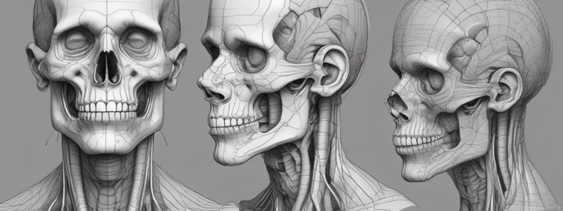
Anatomical Position
- Standard reference position of the body
- Used to describe the location of structures
- Body is upright, with feet together, hands by the side, and face forward
- Mouth is closed, and facial expression is neutral
Anatomical Planes
- Imaginary flat surfaces that traverse the body
- Median plane: divides the body into equal right and left halves
- Sagittal plane: parallel to the median plane
- Frontal (coronal) plane: vertical plane that divides the body into anterior and posterior sections
- Horizontal (transverse) plane: horizontal plane that divides the body into superior and inferior sections
Terms to Describe Location
- Anterior (front) and posterior (back)
- Medial (towards the median) and lateral (away from the median)
- Superior (towards the head) and inferior (towards the feet)
- Proximal (closer to the trunk) and distal (away from the trunk)
- Cranial (towards the head) and caudal (towards the tail)
Anatomical Terms of Movements
- Flexion: decreasing the angle between two structures
- Extension: increasing the angle between two structures
- Abduction: moving away from the midline
- Adduction: moving towards the midline
- Protrusion: moving forward
- Retraction: moving backward
- Depression: moving downward
- Elevation: moving upward
- Medial (internal) rotation: spiral movement towards the midline### Anatomical Terms of Movements
- Lateral (external) rotation: Spiral movement away from the midline
- Inversion: The plantar side of the foot is rotated in the direction of the median plane
- Eversion: The foot is rotated away from the median plane on the plantar side
- Protraction: Moving forwards and laterally simultaneously
- Retrusion: Moving backward (movement of tongue and mandible)
- Flexion (trunk): Side (lateral flexion) or forward (anterior flexion) bending
- Extension (trunk): Bending backward
- Pronation: The radius is rotated medially, causing the palm to face posteriorly (if in anatomical position) or inferiorly (if the elbow is bent)
- Supination: Lateral rotation of the radius, resulting in the palm facing anteriorly (if the elbow is in an anatomical position) or superiorly (if the elbow is flexed)
- Circumduction: A coordinated movement that begins with flexion and progresses through abduction, extension, and adduction
- Deviation: The wrist joint can move in either direction (radial deviation or ulnar deviation)
- Opposition: Using the thumb of the same hand to touch the pad of any of the fingers
- Reposition: Separating any of the fingers' pads from the thumb of the same hand
- Rotation (trunk): Twisting motion towards or away from the midline (left or right)
Surface Landmarks
- Surface landmarks are distinct structures that can be felt or seen directly and remain consistent across individuals
- Examples of surface landmarks:
- Clavicle
- Jugular notch
- Manubrium of sternum
- Sternal angle (2nd costal cartilage)
- Body of sternum
- Xiphoid process of sternum
- Nipple (4th intercostal space)
- Ribs of the pectoral region
- Surface landmarks can be identified in areas such as the abdomen or thorax, including foramina for neurovascular bundles, suture intersections, and bony structures
Anatomical Regions
- All anatomical regions are identified by accurate landmarks, resulting in universally accepted terms
- Regions of the Head and Neck:
- Frontal
- Orbital
- Infraorbital
- Nasal
- Oral
- Mental
- Sternocleidomastoid
- Lateral cervical
- Posterocervical
- Buccal
- Parotideomasseteric
- Infratemporal
- Zygomatic
- Temporal
- Occipital
- Parietal
- Regions of the Lower Extremity:
- Hip
- Gluteal
- Thigh
- Knee
- Leg
- Ankle
- Foot
- Femoral triangle
- Gluteal
- Femoral (anterior, posterior)
- Genicular (anterior, posterior)
- Popliteal
- Crural (anterior, posterior)
- Lateral retromalleolar
- Dorsal
- Plantar
- Calcaneal
Anatomical Lines
- Anatomical lines are imaginary lines drawn vertically or horizontally across an upright
- Examples of anatomical lines:
- Line of symmetry
- Sternal line
- Parasternal line
- Midclavicular line
- Anterior/middle and posterior axillary lines
- Paravertebral line
- Scapular line
- Ribs
- Sternum
- Vertebral spine processes
- Clavicle
- Pectoral muscles
- Anatomical lines provide reference points for locating and describing specific regions on the surface of the body
Cavities of the Body
- Most anatomical structures are found within cavities, which are fluid-filled areas
- Membranes and other structures divide these bodily cavities, providing protection, lubrication, and reducing friction during organ movement
- Each cavity contains unique neurovascular structures and organs specific to its location
- Examples of cavities:
- Ventral or anterior cavity (including the thorax, abdomen, and pelvis)
- Dorsal or posterior cavity (including the cranial and spinal cavities)
- Thoracic cavity
- Abdominopelvic cavity
- Cranial cavity
- Vertebral cavity
Osteology
- Characteristics of bone:
- Living tissue
- Supports the body's skeleton
- Complex, rigid, inflexible, hard, vascular, and dynamic connective tissue
- Composed of cells and intercellular matrix substances
- Types of bone cells:
- Osteoblasts
- Osteocytes
- Osteoclasts
- Bone growth occurs through surface accretions, also known as appositional growth
- Bone function:
- Providing the skeletal frame and body architecture
- Preventing the collapse of the body
- Protecting internal organs from impact-related injuries
- Facilitating the movement of body parts
- Supporting blood formation and the generation of red and white blood cells
- Participating in the buildup and release of substances and minerals
Bone Ossification
- There are two varieties of bone ossification:
- Intramembranous ossification
- Endochondral or intracartilaginous ossification
- Intramembranous ossification:
- Directly replaces the membrane with bone without intervening cartilage stages
- Found in the skull vault and several facial bones
- Endochondral ossification:
- Occurs in the long and short bones of the skeleton
- A model of the bone is first laid down in hyaline cartilage derived from connective tissues in the embryo
Skeletal System
- Divided into two groups:
- Axial skeleton (80 bones)
- Appendicular skeleton (126 bones)
- Axial skeleton:
- Cranium (29 bones)
- Vertebral column and thorax (51 bones)
- Appendicular skeleton:
- Upper extremity bones (64 bones)
- Lower extremity bones (62 bones)
Bones' Types and Classification
- There are two types of bone:
- Compact bone
- Spongy (trabecular or cancellous) bone
- Classification of bones based on anatomical shape:
- Long bones
- Short bones
- Flat bones
- Irregular bones
- Sesamoid bones### Scapulae
- Triangular bone located at the 2nd to 7th levels of the ribs
- Connects the clavicle to the humerus
- Has two faces: costal (anterior) and posterior surfaces
- Features three borders: superior, medial, and lateral borders
- Has three angles: superior, inferior, and lateral angles
Posterior Surface of Scapula
- Convex and features a protrusion called the spine of scapula
- Spine of scapula divides into two concave surfaces: supraspinous fossa and infraspinous fossa
- Supraspinous fossa filled by the supraspinatus muscle and infraspinous fossa filled by the infraspinatus muscle
- Deltoid and trapezius muscles attach to the spine of the scapula
Borders of Scapula
- Superior border: shortest and thinnest, with a slight concave shape
- Lateral border: strong and thick, situated closest to the axilla
- Medial border: long, medial edge of the scapula closest to the vertebral column
Angles of Scapula
- Superior angle: formed by the junction of the superior and medial borders
- Inferior angle: formed by the junction of the medial and lateral borders
- Lateral angle: formed by the junction of the superior and lateral borders, contains the head and neck of the scapula
Glenoid Cavity
- Shallow pit on the lateral margin of the scapula's head, forms the glenohumeral joint with the head of the humerus
- Above the glenoid cavity: a small ridge called the supraglenoid tubercle, where the long head of the biceps brachii muscle attaches
- Below the glenoid cavity: a roughened area known as the infraglenoid tubercle, where the long head of the triceps brachii muscle attaches
Acromion and Coracoid Process
- Acromion: broad, straight lateral extension of the spine of scapula, articulates with the clavicle at the acromioclavicular joint
- Coracoid process: thick, beak-like structure that projects anterolaterally from the lateral end of the scapula
Humerus
- Longest bone in the upper limb, located in the arm (brachium)
- Divided into three main regions: proximal extremity, body, and distal extremity
Proximal Extremity of Humerus
- Consists of several structures, including the head, anatomical neck, greater and lesser tubercles, surgical neck, and the upper half of the body
- Head of humerus: rounded and articulates with the glenoid cavity
- Lesser tubercle: small, jagged ridge below the head, situated at the anterior proximal end of the humerus, and medial to the greater tubercle
Body of Humerus
- Also known as the body of the humerus, is the middle portion of the bone
- Features deltoid tuberosity, a rough area on the lateral side of the body where the deltoid attaches
Distal Extremity of Humerus
- Known as the condyle, located on the lateral side of the distal end of the humerus
- Spherical capitulum: articulates with the head of the radius
- Trochlea: roller-shaped, articulates with the ulna
- Medial and lateral epicondyles: knob-like prominences on the distal, medial and lateral sides of the humerus
Clinical Correlation
- Fractures of the humerus: can lead to direct injury or compression of the axillary nerve, radial nerve, and median and ulnar nerves
- Medial epicondyle fracture: can result in ulnar nerve injuries
- Lateral epicondyle fracture: can result in radial nerve injuries
Radius
- Bone located on the lateral side of the forearm (antebrachium)
- Responsible for pronation and supination movements
- Articulates with the humerus, radius, scaphoid, and lunate bones
- Upper end of the radius: the proximal extremity, is the thickest and strongest part
- Head of the radius: articulates proximally with both the humeral capitulum and the radial notch of the ulna at the elbow joint
Ulna
- Bone located on the medial side of the forearm (antebrachium)
- Articulates with the radius and humerus
- Does not have direct contact with the wrist bones; instead, there is a disc between them
- Upper end of the ulna: the proximal extremity, is the thickest and strongest part
- Olecranon: elbow protrusion, forms the uppermost part of the ulna, and can be easily palpated
Bones of the Hand
- The hand skeleton consists of 27 bones and can be divided into three groups: carpal bones (8), metacarpals (5), and phalanges (14)
- Carpal bones: eight small bones that form the wrist
- Metacarpals: five bones in each hand, each with a head, body, and base from proximal to distal
- Phalanges: 14 bones in each hand, with two in the thumb and three in the other fingers
Studying That Suits You
Use AI to generate personalized quizzes and flashcards to suit your learning preferences.
Related Documents
Description
Discover the tips and strategies to succeed in anatomy studies, including effective learning methods, preparation techniques, and utilizing various resources at Altınbas University.


