30 Questions
What is the weakest part of the humerus bone?
Surgical neck
Which muscle inserts into the crest of the intertubercular groove?
Pectoralis major
What is the name of the artery that runs alongside the radial nerve in the sulcus?
Profunda brachii artery
What is the name of the bony prominence on the lateral side of the humerus where the deltoid muscle attaches?
Deltoid tuberosity
What is the name of the medial bony prominence at the distal end of the humerus?
Medial epicondyle
What is the name of the lateral bony prominence at the distal end of the humerus that articulates with the head of the radius?
Capitulum
What is the characteristic deformity resulting from a Colles fracture?
Dinner fork deformity
Which of the following structures is located on the medial side of the forearm?
Ulna
What is the function of the dorsal tubercle on the posterior face of the radius?
To provide attachment for muscles that extend to the fingers
What is the articular surface on the inner side of the lower end of the radius called?
Ulnar notch
What is the thickest and strongest part of the ulna?
Proximal extremity
What is the direction of the fracture line in a Colles fracture?
Transverse or oblique
What is the function of the radial fossa and coronoid fossa?
To provide a greater range of motion to the forearm
Which nerve can be injured in a medial epicondyle fracture?
Ulnar nerve
What is the term for the depression on the posterior surface of the humerus near the lower end?
Olecranon fossa
Which muscles attach to the greater and lesser tubercles?
Rotator cuff muscles
What can result from a shaft fracture of the humerus?
Direct trauma or compression of the radial nerve
What can result from a distal part fracture of the humerus?
Direct trauma or compression of the median and ulnar nerves and the brachial artery
Which carpal bone has a hook-shaped projection on the palmar side?
Hamate
Which of the following bones articulates with the scaphoid and the first metacarpal bone?
Trapezium
How many bones does the trapezoid articulate with?
Four
Which of the following tendons attaches to the pisiform bone?
Flexor carpi ulnaris
Which carpal bone is located in the center of the wrist?
Capitate
Which of the following bones articulates with the lunate and scaphoid bones?
Capitate
How many bones are in the hand skeleton?
27
Which carpal bone is most frequently fractured?
Scaphoid
Which bone articulates with the radius proximally, the trapezium and trapezoid distally, and the lunate and capitate medially?
Scaphoid
Which bone has no direct contact with the ulna?
Triquetrum
How many bones are in the distal row of the carpal bones?
4
What is the name of the ridge on the palmar surface of the scaphoid bone?
Tubercle
Study Notes
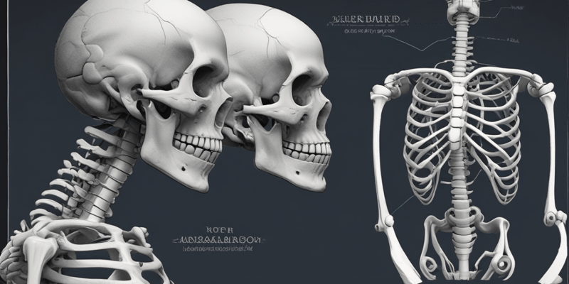
Humerus
- The surgical neck is the weakest part of the bone and can be susceptible to fractures, which may damage the axillary nerve, circumflex humeral artery, and vein.
- The crest of intertubercular groove is a sharp edge where the pectoralis major muscle inserts, while the crest of lesser tubercle is another sharp edge where the teres major inserts.
- The humeral body features a deltoid tuberosity, a rough area on the lateral side of the body where the deltoid attaches.
- The posterior edge of the tuberosity forms a groove (sulcus) for the radial nerve, which courses together with the profunda brachii artery (deep artery of the arm) and can be vulnerable to injury in cases of humerus fractures.
- The medial and lateral supracondylar ridges are sharp edges where the fascia and muscles attach, extending to the medial and lateral epicondyles at the distal end.
Humerus Distal Extremity
- The distal end of the humerus is known as the condyle, featuring a spherical capitulum that articulates with the head of radius, and a roller-shaped trochlea that articulates with the ulna.
- The olecranon fossa is a significant depression on the posterior surface near the lower end, where the olecranon (projection of the ulna) fits into.
- The radial fossa is located on the anterior-superior side of the capitulum, while the coronoid fossa is located anteriorly to the trochlea, providing a greater range of motion to the forearm.
Clinical Correlation: Fractures of the Humerus
- Surgical neck fractures can lead to direct injury or compression of the axillary nerve, resulting in functional impairment of the shoulder joint, specifically affecting abduction and external rotation.
- Shaft fractures can result in direct trauma or compression of the radial nerve, leading to functional impairment of the forearm and hand, specifically affecting extension at the elbow and wrist joints.
- Distal part fractures are rare but can result in direct trauma or compression of the median and ulnar nerves and the brachial artery.
Radius
- The distal extremity of the radius is wider than the other parts and features a raised area called the dorsal tubercle, where the tendons of the muscles that extend to the fingers sit.
- The radial styloid process is a projection on the outer side of the lower end, extending downward.
- The ulnar notch is a crescent-shaped articular surface on the inner side, articulating with the articular circumference of the ulna.
Clinical Correlation: Radius
- Colles fracture is a common type of fracture involving the distal end of the radius, typically occurring as a result of a fall onto an outstretched hand.
- The fracture involves a break in the radius near the wrist joint, specifically at the distal metaphysis or just above it, with the distal fragment displaced dorsally, resulting in a characteristic dinner fork deformity.
Ulna
- The ulna is located on the medial side of the forearm, articulating with the radius and humerus.
- It does not have direct contact with the wrist bones; instead, there is a disc between them.
Carpal Bones
- The carpal bones are eight small bones that form the wrist, arranged in two rows: proximal and distal.
- The proximal row consists of the scaphoid, lunate, triquetrum, and pisiform, while the distal row consists of the trapezium, trapezoid, capitate, and hamate.
Bones of the Proximal Row
- Scaphoid: the largest bone in the proximal row, most frequently fractured, articulates with the radius proximally, the trapezium and trapezoid distally, and the lunate and capitate medially.
- Lunate: a crescent-shaped bone, dislocation of which can result in median nerve damage, articulates with five bones: the radius proximally, the capitate and hamate distally, the scaphoid laterally, and the triquetrum medially.
- Triquetrum: located on the ulnar side of the proximal row, articulates with the ulna through the lunate laterally, the pisiform anteriorly, the hamate distally, and the articular disc proximally.
- Pisiform: the smallest of the carpal bones, located in front of the other bones, articulates with the triquetrum dorsally.
Bones of the Distal Row
- Trapezium: located on the radial side of the wrist, articulates with the scaphoid proximally, the first metacarpal distally, and the trapezoid and second metacarpal bones medially.
- Trapezoid: the smallest bone in the distal row, articulates with four bones: the scaphoid proximally, the second metacarpal distally, the trapezium laterally, and the capitate medially.
- Capitate: the largest of the carpal bones, located in the center of the wrist, articulates with the lunate and scaphoid proximally, the second, third, and fourth metacarpal bones distally, the trapezoid laterally, and the hamate medially.
- Hamate: located on the inferomedial part of the wrist, articulates with the lunate proximally, the fourth and fifth metacarpal bones distally, the triquetrum medially, and the capitate laterally.
This quiz covers the structure and features of the humerus bone in the human body, including its weakest parts, muscle insertions, and blood vessel relationships.
Make Your Own Quizzes and Flashcards
Convert your notes into interactive study material.
Get started for free

