Podcast
Questions and Answers
Which bones form the knee joint?
Which bones form the knee joint?
What is the role of the fibula in the knee joint?
What is the role of the fibula in the knee joint?
Which structures articulate at the ankle joint?
Which structures articulate at the ankle joint?
What forms the pelvic girdle?
What forms the pelvic girdle?
Signup and view all the answers
Which of the following bones does not articulate with the tibia?
Which of the following bones does not articulate with the tibia?
Signup and view all the answers
What is the origin of the gluteus medius muscle?
What is the origin of the gluteus medius muscle?
Signup and view all the answers
Where does the gluteus minimus insert?
Where does the gluteus minimus insert?
Signup and view all the answers
Which nerve innervates the gluteus medius muscle?
Which nerve innervates the gluteus medius muscle?
Signup and view all the answers
What distinguishes the gluteus minimus from the gluteus medius?
What distinguishes the gluteus minimus from the gluteus medius?
Signup and view all the answers
Which muscle is not considered a lateral rotator of the hip?
Which muscle is not considered a lateral rotator of the hip?
Signup and view all the answers
What is the main functional purpose of the pelvic ligaments?
What is the main functional purpose of the pelvic ligaments?
Signup and view all the answers
Which part of the coxal bone is primarily responsible for forming the hip joint with the femur?
Which part of the coxal bone is primarily responsible for forming the hip joint with the femur?
Signup and view all the answers
What are the fused vertebrae that comprise the sacrum?
What are the fused vertebrae that comprise the sacrum?
Signup and view all the answers
Which landmark is NOT found on the ischium of the coxal bone?
Which landmark is NOT found on the ischium of the coxal bone?
Signup and view all the answers
What are the three smaller bones that fuse to form each coxal bone?
What are the three smaller bones that fuse to form each coxal bone?
Signup and view all the answers
What is the primary action facilitated by the gluteus medius muscle at the hip?
What is the primary action facilitated by the gluteus medius muscle at the hip?
Signup and view all the answers
Which of the following muscles is NOT innervated by the superior gluteal nerve?
Which of the following muscles is NOT innervated by the superior gluteal nerve?
Signup and view all the answers
What anatomical feature distinguishes the gluteus minimus from the gluteus medius?
What anatomical feature distinguishes the gluteus minimus from the gluteus medius?
Signup and view all the answers
Which of the following muscles is primarily responsible for lateral rotation of the hip?
Which of the following muscles is primarily responsible for lateral rotation of the hip?
Signup and view all the answers
What anatomical feature differentiates the left femur from the right femur when using the 'femur gun' method?
What anatomical feature differentiates the left femur from the right femur when using the 'femur gun' method?
Signup and view all the answers
Which muscle originates from the gluteal fossa?
Which muscle originates from the gluteal fossa?
Signup and view all the answers
Which implication does a larger Q-angle have in females compared to males?
Which implication does a larger Q-angle have in females compared to males?
Signup and view all the answers
What contributes to the stability of the hip joint compared to the shoulder joint?
What contributes to the stability of the hip joint compared to the shoulder joint?
Signup and view all the answers
What type of injuries are commonly associated with the hip joint?
What type of injuries are commonly associated with the hip joint?
Signup and view all the answers
Which ligaments are involved in the structure of the hip joint?
Which ligaments are involved in the structure of the hip joint?
Signup and view all the answers
What does the tibia articulate with at the ankle joint?
What does the tibia articulate with at the ankle joint?
Signup and view all the answers
Which joint connects the left and right coxal bones?
Which joint connects the left and right coxal bones?
Signup and view all the answers
Which structure is adapted to provide weightbearing and stability in the knee?
Which structure is adapted to provide weightbearing and stability in the knee?
Signup and view all the answers
Which of the following bones does not form part of the pelvic girdle?
Which of the following bones does not form part of the pelvic girdle?
Signup and view all the answers
What articulates with the inferior aspect of the sacrum?
What articulates with the inferior aspect of the sacrum?
Signup and view all the answers
What is the insertion point for the gemellus inferior muscle?
What is the insertion point for the gemellus inferior muscle?
Signup and view all the answers
Which of the following muscles passes through the lesser sciatic foramen?
Which of the following muscles passes through the lesser sciatic foramen?
Signup and view all the answers
How does the action of the piriformis muscle differ from that of the quadratus femoris muscle?
How does the action of the piriformis muscle differ from that of the quadratus femoris muscle?
Signup and view all the answers
Which nerve innervates the gemellus superior muscle?
Which nerve innervates the gemellus superior muscle?
Signup and view all the answers
Which structure primarily provides stability to the hip joint compared to other joints in the body?
Which structure primarily provides stability to the hip joint compared to other joints in the body?
Signup and view all the answers
What is a function of the intrinsic muscles of the hip and gluteal region?
What is a function of the intrinsic muscles of the hip and gluteal region?
Signup and view all the answers
Which of the following pairs of bones articulate to form the hip joint?
Which of the following pairs of bones articulate to form the hip joint?
Signup and view all the answers
Which of the following statements accurately describes the coxal bone structure?
Which of the following statements accurately describes the coxal bone structure?
Signup and view all the answers
Predict the functional implication of an injury to the superior gluteal nerve.
Predict the functional implication of an injury to the superior gluteal nerve.
Signup and view all the answers
What is the primary innervation of both the gluteus medius and gluteus minimus muscles?
What is the primary innervation of both the gluteus medius and gluteus minimus muscles?
Signup and view all the answers
What is the primary structural component of the coxal bone?
What is the primary structural component of the coxal bone?
Signup and view all the answers
What distinguishes the gluteus minimus from the gluteus medius in terms of anatomical position?
What distinguishes the gluteus minimus from the gluteus medius in terms of anatomical position?
Signup and view all the answers
Which ligament is primarily responsible for stabilizing the sacroiliac joint?
Which ligament is primarily responsible for stabilizing the sacroiliac joint?
Signup and view all the answers
Which muscle can be classified as a lateral hip rotator situated near the gluteal region?
Which muscle can be classified as a lateral hip rotator situated near the gluteal region?
Signup and view all the answers
Which landmark is closely associated with the ischium?
Which landmark is closely associated with the ischium?
Signup and view all the answers
Where does the gluteus medius muscle primarily insert?
Where does the gluteus medius muscle primarily insert?
Signup and view all the answers
Which statement accurately describes the action of the gluteus medius muscle?
Which statement accurately describes the action of the gluteus medius muscle?
Signup and view all the answers
What anatomical feature distinguishes the hip joint from other joints such as the shoulder?
What anatomical feature distinguishes the hip joint from other joints such as the shoulder?
Signup and view all the answers
Which of the following is true regarding the pelvis and its ligaments?
Which of the following is true regarding the pelvis and its ligaments?
Signup and view all the answers
Which artery is primarily responsible for supplying the posterior compartment of the thigh?
Which artery is primarily responsible for supplying the posterior compartment of the thigh?
Signup and view all the answers
What is the primary source of blood supply to the head of the femur?
What is the primary source of blood supply to the head of the femur?
Signup and view all the answers
Which artery supplies the medial compartment of the thigh?
Which artery supplies the medial compartment of the thigh?
Signup and view all the answers
Which artery branches from the descending aorta to supply the gluteal region?
Which artery branches from the descending aorta to supply the gluteal region?
Signup and view all the answers
Which artery does NOT branch from the femoral artery to supply the different compartments of the leg?
Which artery does NOT branch from the femoral artery to supply the different compartments of the leg?
Signup and view all the answers
Flashcards
Femur articulation
Femur articulation
The femur joins the patella and tibia to form the knee joint.
Fibula articulation
Fibula articulation
The fibula connects to the tibia, contributing to the ankle joint.
Ankle joint bones
Ankle joint bones
The tibia, fibula, and tarsal bones meet to create the ankle joint.
Pelvic girdle components
Pelvic girdle components
Signup and view all the flashcards
Coccyx articulation
Coccyx articulation
Signup and view all the flashcards
Sacrum
Sacrum
Signup and view all the flashcards
Coxal bone
Coxal bone
Signup and view all the flashcards
Pelvic girdle
Pelvic girdle
Signup and view all the flashcards
Hip joint
Hip joint
Signup and view all the flashcards
Femur
Femur
Signup and view all the flashcards
Gluteus Medius Muscle Origin
Gluteus Medius Muscle Origin
Signup and view all the flashcards
Gluteus Medius Muscle Insertion
Gluteus Medius Muscle Insertion
Signup and view all the flashcards
Gluteus Medius Nerve Supply
Gluteus Medius Nerve Supply
Signup and view all the flashcards
Gluteus Minimus Muscle Origin
Gluteus Minimus Muscle Origin
Signup and view all the flashcards
Gluteus Minimus Muscle Insertion
Gluteus Minimus Muscle Insertion
Signup and view all the flashcards
What bones form the knee joint?
What bones form the knee joint?
Signup and view all the flashcards
What is the function of the fibula at the knee?
What is the function of the fibula at the knee?
Signup and view all the flashcards
What bones form the ankle joint?
What bones form the ankle joint?
Signup and view all the flashcards
What forms the pelvic girdle?
What forms the pelvic girdle?
Signup and view all the flashcards
Where does the coccyx connect?
Where does the coccyx connect?
Signup and view all the flashcards
Femur Gun
Femur Gun
Signup and view all the flashcards
Hip Joint Movements
Hip Joint Movements
Signup and view all the flashcards
Sex Differences in Hips
Sex Differences in Hips
Signup and view all the flashcards
Iliofemoral, Pubofemoral, Ischiofemoral Ligaments
Iliofemoral, Pubofemoral, Ischiofemoral Ligaments
Signup and view all the flashcards
Gluteus Medius Function
Gluteus Medius Function
Signup and view all the flashcards
Gluteus Medius Origin
Gluteus Medius Origin
Signup and view all the flashcards
Gluteus Medius Insertion
Gluteus Medius Insertion
Signup and view all the flashcards
Gluteus Minimus Function
Gluteus Minimus Function
Signup and view all the flashcards
Gluteus Minimus Origin
Gluteus Minimus Origin
Signup and view all the flashcards
Lateral Hip Rotators
Lateral Hip Rotators
Signup and view all the flashcards
Piriformis Muscle
Piriformis Muscle
Signup and view all the flashcards
Quadratus Femoris Muscle
Quadratus Femoris Muscle
Signup and view all the flashcards
Obturator Internus Muscle
Obturator Internus Muscle
Signup and view all the flashcards
Gemellus Superior & Inferior Muscles
Gemellus Superior & Inferior Muscles
Signup and view all the flashcards
Hip Joint Formation
Hip Joint Formation
Signup and view all the flashcards
What are the primary functions of the gluteus medius and minimus?
What are the primary functions of the gluteus medius and minimus?
Signup and view all the flashcards
What are the three bones that fuse to form the coxal bone?
What are the three bones that fuse to form the coxal bone?
Signup and view all the flashcards
What is the acetabulum?
What is the acetabulum?
Signup and view all the flashcards
What are the major landmarks on the proximal femur?
What are the major landmarks on the proximal femur?
Signup and view all the flashcards
What is the pubic symphysis?
What is the pubic symphysis?
Signup and view all the flashcards
What are the sacroiliac joints?
What are the sacroiliac joints?
Signup and view all the flashcards
Gluteus Medius & Minimus Origin
Gluteus Medius & Minimus Origin
Signup and view all the flashcards
What are perforating branches?
What are perforating branches?
Signup and view all the flashcards
What compartment does the femoral artery supply primarily?
What compartment does the femoral artery supply primarily?
Signup and view all the flashcards
What does the obturator artery supply?
What does the obturator artery supply?
Signup and view all the flashcards
What is the main branch of the posterior tibial artery?
What is the main branch of the posterior tibial artery?
Signup and view all the flashcards
What structure does the gluteal artery branch from?
What structure does the gluteal artery branch from?
Signup and view all the flashcards
Study Notes
Western University, Canada
- Western University, Canada is a prominent educational institution.
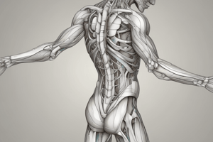
Hip and Gluteal Region
- The hip and gluteal region is a complex anatomical area.
- Learning outcomes include identifying bones and ligaments, explaining hip movements, identifying and describing intrinsic hip/gluteal muscles, understanding innervations, and predicting implications of musculoskeletal injuries.
Regions of the Lower Limb
- The lower limb is divided into regions including the hip, thigh, knee, leg, and foot.
Bones of the Lower Limb
- The coxal (hip) bone is formed from three bones (ilium, ischium, pubis).
- The left and right coxal bones articulate at the pubic symphysis and sacroiliac joints to form the pelvic girdle.
- The coxal bone connects with the femur at the hip joint.
- The femur articulates with the patella and tibia.
- The fibula articulates with the tibia and tarsal bones at the ankle joint.
Bones, Joints, and Ligaments
- This section focuses on the structural elements of the hip and pelvic area.
Bones of the Hip and Pelvis
- The left and right coxal bones articulate with the sacrum posteriorly (sacroiliac joints) and anteriorly (pubic symphysis), creating the pelvic girdle.
- The coccyx connects to the inferior aspect of the sacrum.
- The femur connects with the hip joint
Coxal Bone: Ilium
- The ilium is a significant part of the coxal bone.
- The ilium has bony features, including the iliac crest, posterior and anterior superior/inferior iliac spines (PSIS/PIIS/ASIS/AIIS).
- These serve as spatial landmarks and attachment points for muscles.
Coxal Bone: Ischium
- The ischium's key landmarks are the ischial spine and tuberosity, which are important muscle attachments.
- The greater and lesser sciatic notches are important neurovascular passageways.
Coxal Bone: Pubis
- The pubic bone is part of the coxal bone.
- The pubic bones articulate with each other at the pubic symphysis.
- Key landmarks include the superior and inferior pubic rami, the pubic tubercle, and the obturator foramen.
Pelvic Joints and Ligaments
- Pelvic girdle stability relies on ligaments.
- Lumbosacral and sacroiliac joints are crucial pelvic areas.
- Traumatic events (childbirth, accidents) can cause ligament tears, potentially leading to separation.
Pelvic Ligaments
- Pelvic ligaments act as landmarks for muscles, and help stabilize joints.
Hip Joint and Proximal Femur
- The head of the femur connects with the acetabulum of the coxal bone to form the hip joint.
- Parts of the proximal femur are landmarks for attachment of muscles.
Surface Anatomy
- Surface landmarks on the coxal bone and femur, which are palpable, help with clinical assessments and interventions.
The 'Femur Gun'
- Anatomical features of the femur are compared to a handgun.
- The parts of the femur are "hammer", "grip", "trigger" etc.
Hip Joint
- The hip joint, like the shoulder joint, is a ball-and-socket joint.
Movements at the Hip
- The hip, as a ball-and-socket joint, has a wide range of motion, including flexion, extension, abduction, adduction, rotation, and circumduction.
Hip Joint and Femur: Sex Differences
- Anatomical differences in hip angles, such as the Q-angle, exist between sexes.
- These differences relate to clinical considerations.
Hip Joint and Ligaments
- Ligaments of the hip, such as the iliofemoral, pubofemoral, and ischiofemoral ligaments, have a spiral appearance due to lower limb development.
Hip Joint Injury
- Common hip injuries include dislocation and femoral neck fractures.
Gluteal Region Muscles
- Detailed information on muscles is provided, but memorization is not required.
- A muscle chart is available.
Fascia
- Tissues, such as the iliotibial band, which is a type of fascial thickening, encases muscles in the thigh and separates them into compartments.
Superficial Gluteal Muscles
- This section describes the major superficial gluteal muscles, including origin, insertion, innervation, and function.
Deep Gluteal Muscles
- Anatomical details, like origins, insertions, and functions are presented for deep gluteal muscles.
Lateral Hip Rotator Muscles
- The anatomy of muscles that contribute to hip lateral rotation are described.
- The uniqueness of the obturator internus muscle is highlighted.
Lateral Hip Rotator Muscles 2
- The details of the gemellus superior and inferior muscles are covered.
Lateral Hip Rotator Muscles 3
- The location of obturator externus muscle is explained.
Neurovasculature
- This section focuses on the blood supply (arteries and veins) to the lower limb and the nervous system elements..
Arteries of the Lower Limb
- The arterial supply of the lower limb is discussed, including details on branches and perforating vessels.
Gluteal Region Arteries
- The blood supply to the gluteal area (from branches of major arteries) is explained.
Veins of the Lower Limb
- The venous pathway of the lower limb is presented, emphasizing similarities and differences compared to arteries..
Lumbosacral Plexus
- The lumbar and sacral plexuses, which constitute the lumbosacral plexus, are discussed.
- This plexus innervates the lower limb.
Gluteal Region Nerves
- Nerves that supply the gluteal region—especially the superior and inferior gluteal nerves—are detailed.
Dermatomes vs. Nerve Maps
- The relationship between dermatomes and nerve maps is reviewed.
- The overlapping nature of nerve supply is highlighted.
Simplified Muscle Chart
- A chart summarizes the major hip muscles with information on origin, insertion, function, and nerve supply.
Upper Limb vs. Lower Limb
- Functional differences and similarities between upper and lower limbs are presented in a tabular format.
Learning Outcomes (reiteration)
- Review of the key understandings about the hip and gluteal region is provided in terms of learning objectives.
Studying That Suits You
Use AI to generate personalized quizzes and flashcards to suit your learning preferences.
Related Documents
Description
This quiz focuses on the anatomy of the hip and gluteal region, as well as other regions of the lower limb. Participants will learn about the bones, joints, ligaments, and movements associated with these areas. The outcomes include identifying major structures and understanding the implications of injuries.