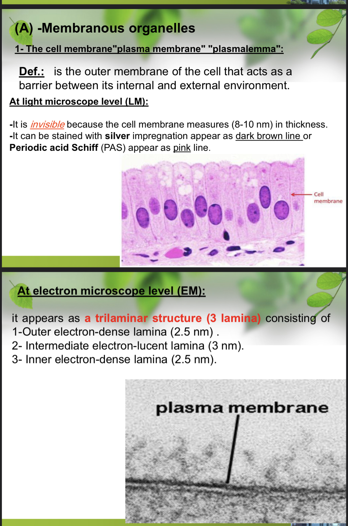
What is the structure and function of the cell membrane at light and electron microscope levels?

Understand the Problem
The question is about the structure and properties of the cell membrane, also known as the plasma membrane, at different levels of microscopy—the light microscope (LM) and electron microscope (EM). It describes how the cell membrane appears and its components at these levels, including its thickness, staining characteristics, and structural layers.
Answer
Invisible at light microscope, trilaminar (3 layers) at electron microscope.
The cell membrane, at the light microscope level, appears invisible due to its thinness (8-10 nm) but can be highlighted with special stains. At the electron microscope level, it shows a trilaminar structure: outer electron-dense lamina (2.5 nm), intermediate electron-lucent lamina (3 nm), and inner electron-dense lamina (2.5 nm).
Answer for screen readers
The cell membrane, at the light microscope level, appears invisible due to its thinness (8-10 nm) but can be highlighted with special stains. At the electron microscope level, it shows a trilaminar structure: outer electron-dense lamina (2.5 nm), intermediate electron-lucent lamina (3 nm), and inner electron-dense lamina (2.5 nm).
More Information
The cell membrane acts as a crucial barrier and gateway for substances entering and leaving the cell, with its trilaminar structure revealed only through electron microscopy.
Tips
A common mistake is to assume the membrane is visible under a light microscope without special staining due to its thinness.
Sources
- Structure of the Plasma Membrane - The Cell - NCBI Bookshelf - ncbi.nlm.nih.gov
- Cell membrane - Wikipedia - en.wikipedia.org
- Looking at the Structure of Cells in the Microscope - NCBI - ncbi.nlm.nih.gov
AI-generated content may contain errors. Please verify critical information