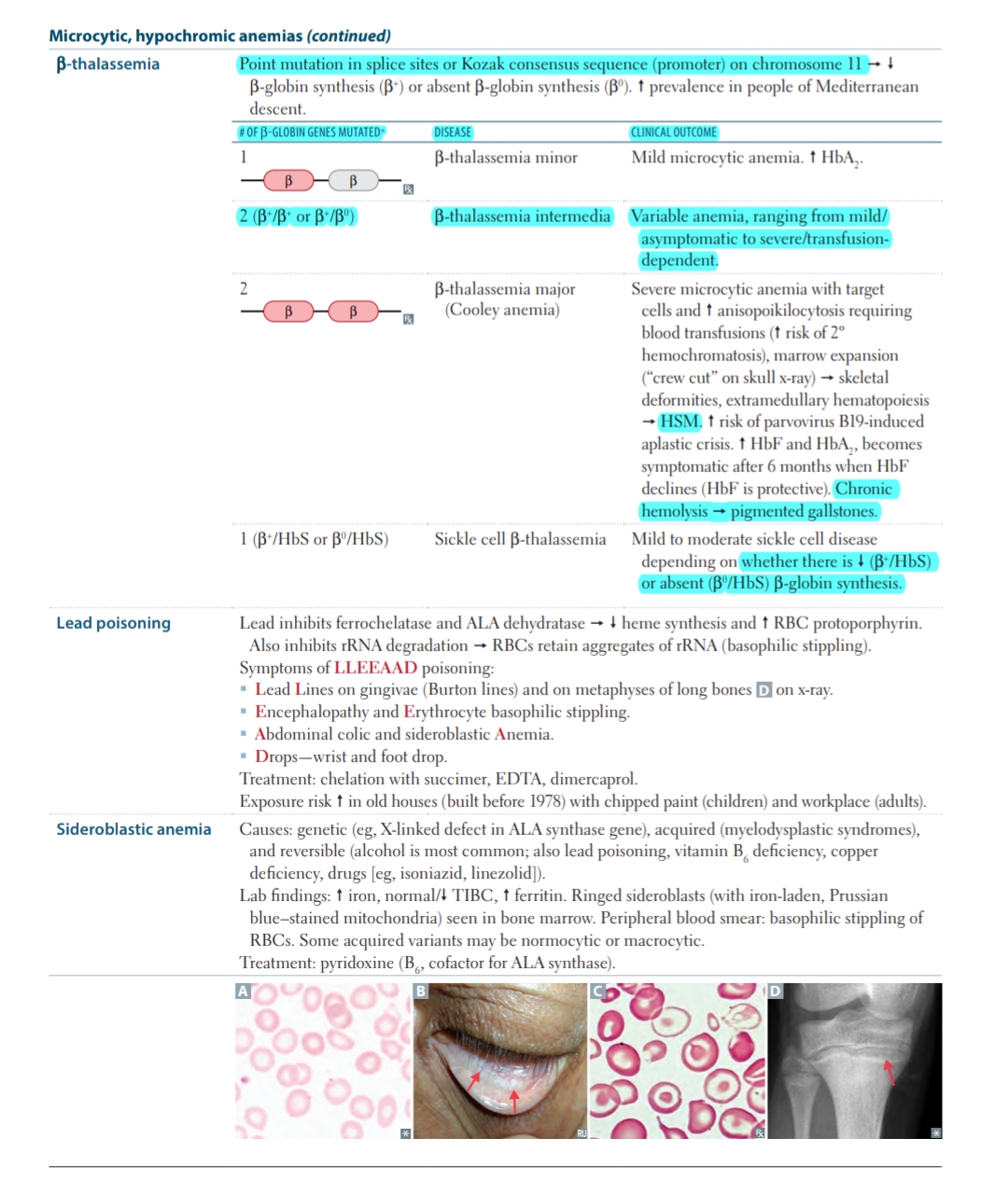
What are the types and characteristics of microcytic, hypochromic anemias, particularly beta-thalassemia, lead poisoning, and sideroblastic anemia?

Understand the Problem
The text provides information about different types of microcytic, hypochromic anemias, particularly focusing on beta-thalassemia, lead poisoning, and sideroblastic anemia, including their genetic causes, clinical outcomes, symptoms, and treatments.
Answer
The types and characteristics of microcytic hypochromic anemias include beta-thalassemia, lead poisoning, and sideroblastic anemia.
The types and characteristics of microcytic hypochromic anemias include the following:
-
Beta-thalassemia: Characterized by mutations that reduce or eliminate β-globin synthesis, leading to mild to severe microcytic anemia. Clinical outcomes vary from mild (β-thalassemia minor) to severe (β-thalassemia major/Cooley anemia) depending on the number and type of gene mutations. Key features include target cells, anisopoikilocytosis, marrow expansion, and increased risk of several complications such as secondary hemochromatosis and spleen enlargement (HSM).
-
Lead poisoning: Lead inhibits enzymes necessary for heme synthesis and RBC protoporphyrin, resulting in basophilic stippling in RBCs. Key symptoms include Burton lines on gums, lead lines on bones, encephalopathy, and abdominal colic. Treatment includes chelation therapy.
-
Sideroblastic anemia: Results from faulty heme synthesis; can be genetic (e.g., X-linked defect in ALA synthase) or acquired (like lead poisoning or certain drugs). Characterized by ringed sideroblasts in bone marrow. Peripheral smear shows basophilic stippling. Commonly corrected with vitamin B6 supplementation.
Answer for screen readers
The types and characteristics of microcytic hypochromic anemias include the following:
-
Beta-thalassemia: Characterized by mutations that reduce or eliminate β-globin synthesis, leading to mild to severe microcytic anemia. Clinical outcomes vary from mild (β-thalassemia minor) to severe (β-thalassemia major/Cooley anemia) depending on the number and type of gene mutations. Key features include target cells, anisopoikilocytosis, marrow expansion, and increased risk of several complications such as secondary hemochromatosis and spleen enlargement (HSM).
-
Lead poisoning: Lead inhibits enzymes necessary for heme synthesis and RBC protoporphyrin, resulting in basophilic stippling in RBCs. Key symptoms include Burton lines on gums, lead lines on bones, encephalopathy, and abdominal colic. Treatment includes chelation therapy.
-
Sideroblastic anemia: Results from faulty heme synthesis; can be genetic (e.g., X-linked defect in ALA synthase) or acquired (like lead poisoning or certain drugs). Characterized by ringed sideroblasts in bone marrow. Peripheral smear shows basophilic stippling. Commonly corrected with vitamin B6 supplementation.
More Information
Microcytic hypochromic anemias are characterized by small, pale red blood cells and often involve conditions that interfere with hemoglobin synthesis. Early diagnosis and treatment are crucial in managing these conditions and preventing severe complications.
Tips
Common mistakes include misdiagnosing the type of anemia due to overlapping symptoms and failing to consider genetic history in beta-thalassemia diagnosis.
Sources
- Microcytic Hypochromic Anemia - StatPearls - NCBI Bookshelf - ncbi.nlm.nih.gov
- Beta Thalassemia - StatPearls - NCBI Bookshelf - ncbi.nlm.nih.gov
- Sideroblastic Anemia: Causes, Symptoms & Treatment - my.clevelandclinic.org
AI-generated content may contain errors. Please verify critical information