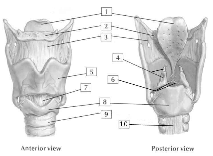
What are the labeled parts in the anatomical diagram of the larynx shown in the image?

Understand the Problem
The question is related to identifying and labeling anatomical parts of the larynx as shown in the provided image. The focus is on understanding the anatomy from both anterior and posterior views.
Answer
1. Epiglottis 2. Hyoid bone 3. Thyrohyoid membrane 4. Thyroid cartilage 5. Cricoid cartilage 6. Arytenoid cartilage 7. Vocal cords 8. Trachea 9. Esophagus 10. Cricoid cartilage (posterior view)
The labeled parts in the anatomical diagram of the larynx are as follows:
- Epiglottis
- Hyoid bone
- Thyrohyoid membrane
- Thyroid cartilage
- Cricoid cartilage
- Arytenoid cartilage
- Vocal cords
- Trachea
- Esophagus
- Cricoid cartilage posterior view
Answer for screen readers
The labeled parts in the anatomical diagram of the larynx are as follows:
- Epiglottis
- Hyoid bone
- Thyrohyoid membrane
- Thyroid cartilage
- Cricoid cartilage
- Arytenoid cartilage
- Vocal cords
- Trachea
- Esophagus
- Cricoid cartilage posterior view
More Information
The larynx, or voice box, is a complex structure involved in breathing, sound production, and protection of the trachea against food aspiration. Its key components include various cartilages such as the thyroid and cricoid, crucial for its function and support.
Tips
Common mistakes can include confusing the thyroid and cricoid cartilages. Remember, the cricoid is ring-shaped and located below the thyroid cartilage.
Sources
- Larynx Anatomy: Image Details - NCI Visuals Online - visualsonline.cancer.gov
- Larynx anatomy: Cartilages, ligaments and muscles | Kenhub - kenhub.com
AI-generated content may contain errors. Please verify critical information