What are the key characteristics and structures of the median cubital vein, cephalic vein, and basilic vein?

Understand the Problem
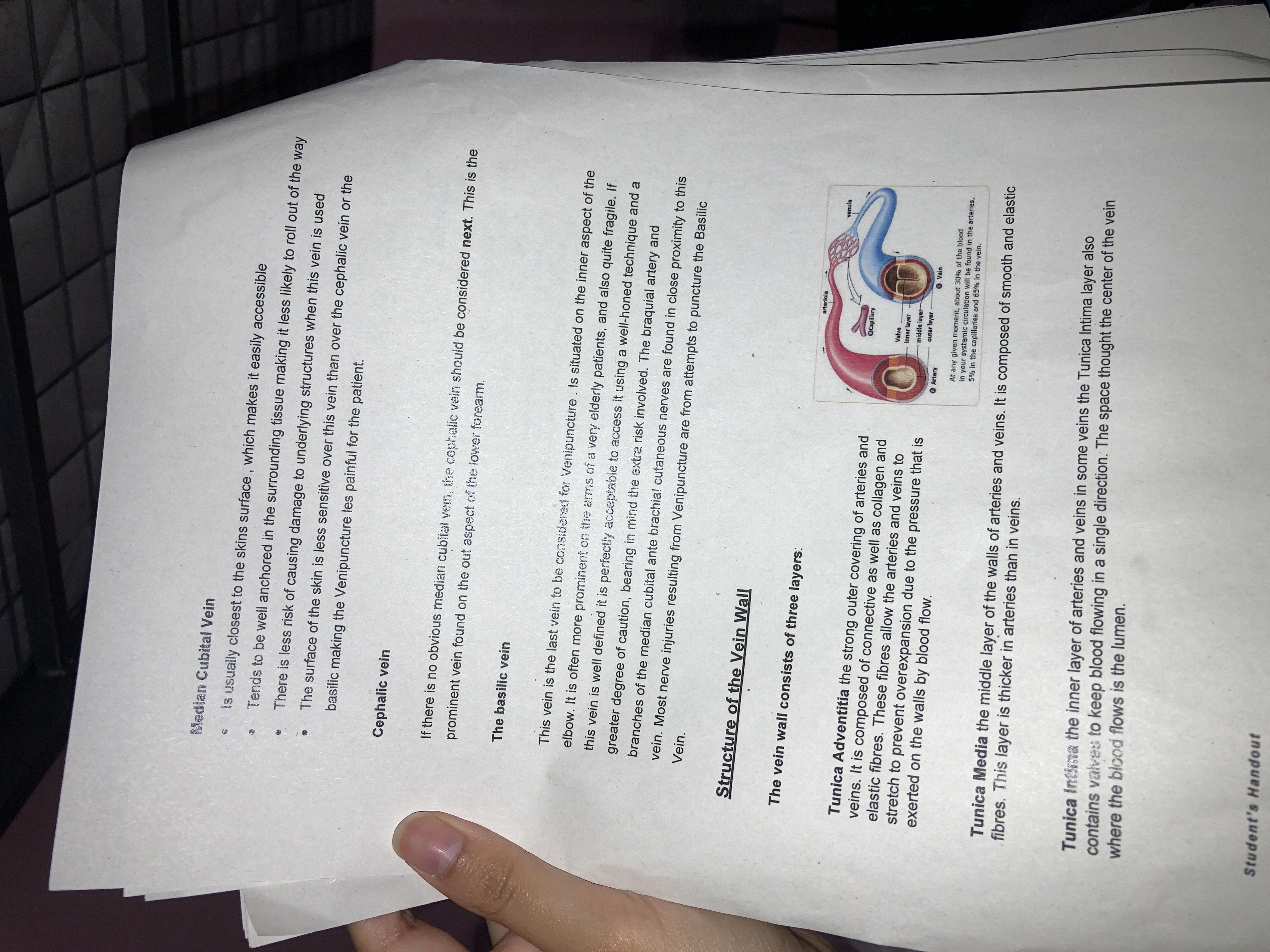
The text provides information about veins used for venipuncture, particularly focusing on the median cubital vein, cephalic vein, and basilic vein, as well as detailing the structure of the vein wall and its layers.
Answer
Median cubital connects basilic and cephalic veins; cephalic is lateral; basilic is medial. Veins have three layers: adventitia, media, intima.
The median cubital vein is a superficial vein in the cubital fossa connecting the basilic and cephalic veins. The cephalic vein is prominent on the lateral forearm. The basilic vein is situated medially. Vein walls have three layers: tunica adventitia, tunica media, and tunica intima.
Answer for screen readers
The median cubital vein is a superficial vein in the cubital fossa connecting the basilic and cephalic veins. The cephalic vein is prominent on the lateral forearm. The basilic vein is situated medially. Vein walls have three layers: tunica adventitia, tunica media, and tunica intima.
More Information
The median cubital vein is often used for venipuncture because it is easily accessible. The cephalic vein is often visible and easily located, providing an additional option for blood draw. The basilic vein is used less frequently due to its deeper position and proximity to nerves.
Tips
Misidentifying the veins during medical procedures like venipuncture can lead to complications. Always verify vein position and use appropriate techniques.
Sources
- Venous Drainage of the Upper Limb - Basilic - teachmeanatomy.info
- Median cubital vein: Anatomy - kenhub.com
- Median Cubital Vein - AnatomyZone - anatomyzone.com
AI-generated content may contain errors. Please verify critical information