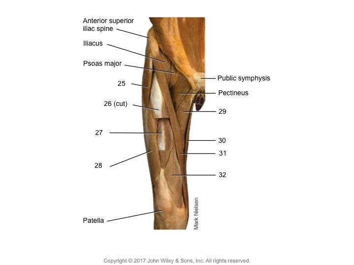
What are the anatomical labels in this image of the dissected leg?

Understand the Problem
The question pertains to the anatomical labels and structures depicted in the provided image of a dissected leg, focusing on muscle and bone identification.
Answer
25 - Sartorius, 26 (cut) - Rectus femoris, 27 - Vastus lateralis, 28 - Vastus medialis, 29 - Adductor longus, 30 - Gracilis, 31 - Adductor magnus, 32 - Semimembranosus.
The image labels: 25 - Sartorius, 26 (cut) - Rectus femoris, 27 - Vastus lateralis, 28 - Vastus medialis, 29 - Adductor longus, 30 - Gracilis, 31 - Adductor magnus, 32 - Semimembranosus.
Answer for screen readers
The image labels: 25 - Sartorius, 26 (cut) - Rectus femoris, 27 - Vastus lateralis, 28 - Vastus medialis, 29 - Adductor longus, 30 - Gracilis, 31 - Adductor magnus, 32 - Semimembranosus.
More Information
The illustration shows key muscle groups and anatomical landmarks that are vital in understanding lower limb anatomy. These muscles are involved in movements like knee extension and hip flexion.
Tips
A common mistake is confusing muscle placement, such as mixing up the vastus lateralis and vastus medialis, which are on opposite sides of the femur.
Sources
- RCSI - Drawing Cross-section of the leg - English labels - anatomytool.org
- Muscles of Leg (Superficial Dissection): Anterior View - Netter Images - netterimages.com
AI-generated content may contain errors. Please verify critical information