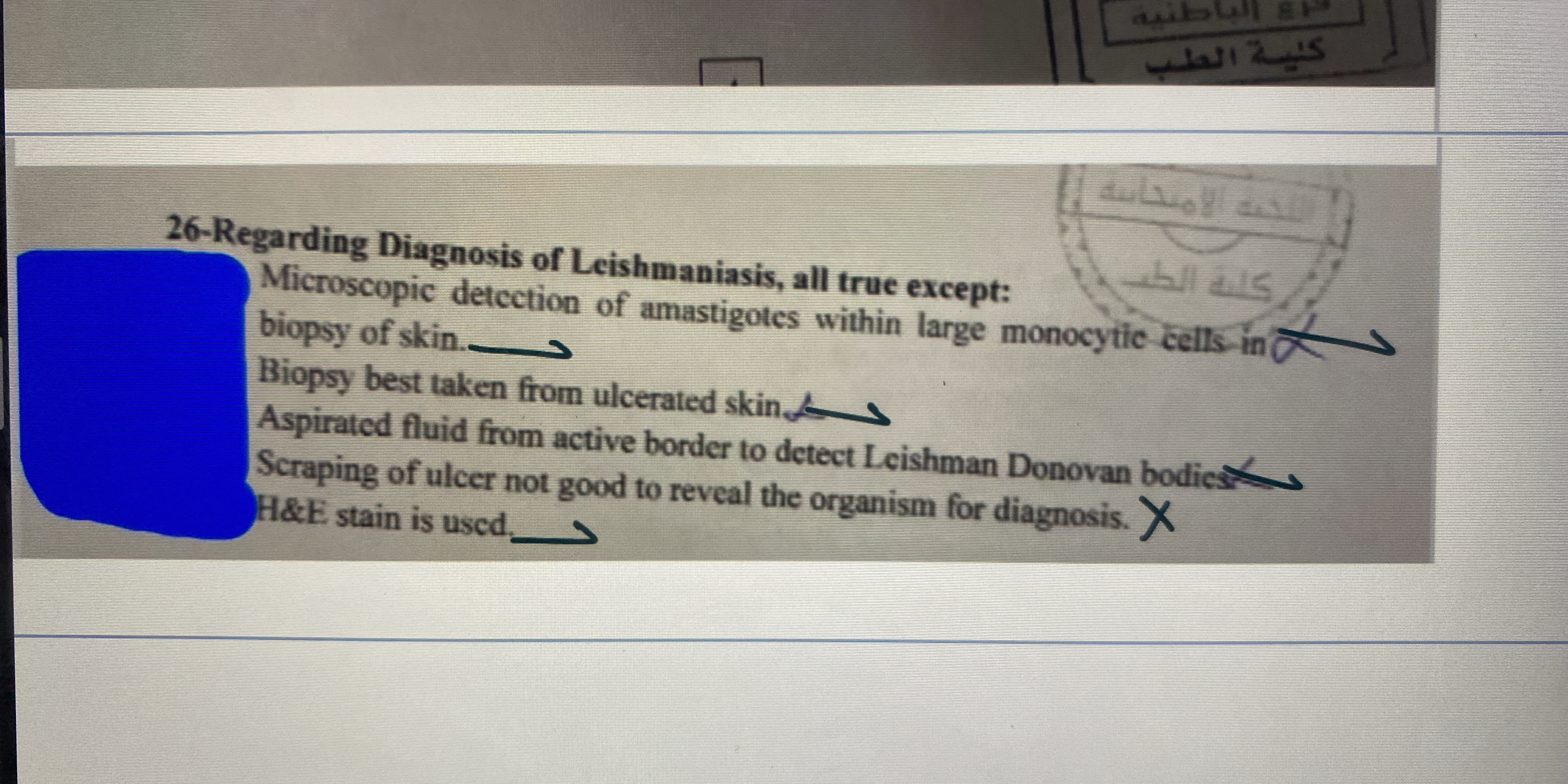
Regarding the Diagnosis of Leishmaniasis, all true except: 1) Microscopic detection of amastigotes within large monocyte cells in biopsy of skin. 2) Biopsy best taken from ulcerate... Regarding the Diagnosis of Leishmaniasis, all true except: 1) Microscopic detection of amastigotes within large monocyte cells in biopsy of skin. 2) Biopsy best taken from ulcerated skin. 3) Aspirated fluid from active border to detect Leishman Donovan bodies. 4) Scraping of ulcer not good to reveal the organism for diagnosis. 5) H&E stain is used.

Understand the Problem
The question asks about the diagnosis of Leishmaniasis, specifically identifying which statement regarding its diagnosis is false. It presents several statements concerning detection methods and requires the respondent to determine the incorrect one.
Answer
Microscopic detection of amastigotes within large monocyte cells in biopsy of skin.
The incorrect statement is: Microscopic detection of amastigotes within large monocyte cells in biopsy of skin.
Answer for screen readers
The incorrect statement is: Microscopic detection of amastigotes within large monocyte cells in biopsy of skin.
More Information
Detection of Leishmania amastigotes typically occurs within macrophages, not specifically large monocyte cells.
Tips
Avoid confusing monocytes with macrophages, which are the actual hosts of Leishmania amastigotes.
Sources
- Diagnosis of Cutaneous Leishmaniasis: Why Punch When You Can ... - pmc.ncbi.nlm.nih.gov
- Isolation of Leishmania organisms - CDC - cdc.gov
- Leishmaniasis Workup - Medscape Reference - emedicine.medscape.com
AI-generated content may contain errors. Please verify critical information