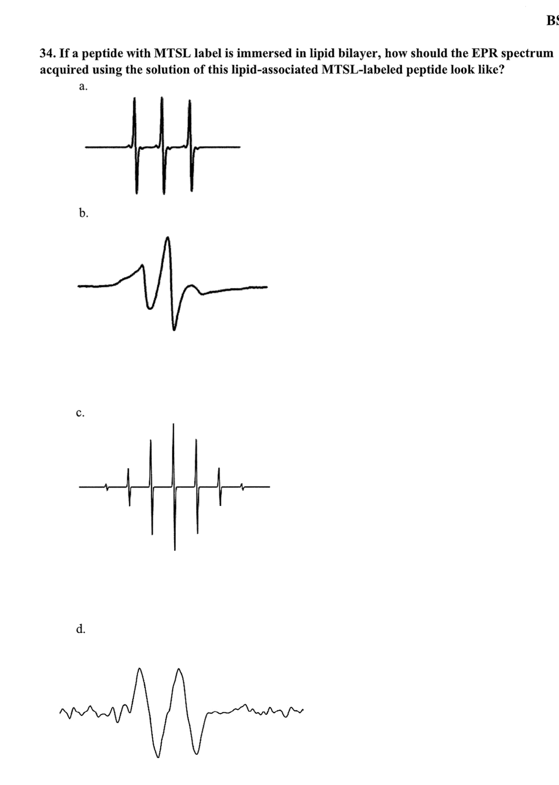
If a peptide with MTSL label is immersed in lipid bilayer, how should the EPR spectrum acquired using the solution of this lipid-associated MTSL-labeled peptide look like?

Understand the Problem
The question is asking how the EPR (Electron Paramagnetic Resonance) spectrum of a peptide with an MTSL label should look when immersed in a lipid bilayer. It presents four options (a, b, c, d) showing different EPR spectra, prompting the user to select the correct representation.
Answer
c
The final answer is c.
Answer for screen readers
The final answer is c.
More Information
In lipid bilayers, EPR spectra of MTSL-labeled peptides often show broadened features due to restricted motion, indicating fewer degrees of freedom compared to the same label in solution.
Tips
One common mistake is to misinterpret motion restriction in lipid environments, leading to incorrect spectral interpretations.
Sources
- Site‐Directed Spin Labeling EPR for Studying Membrane Proteins - onlinelibrary.wiley.com
- Peptide-membrane Interactions by Spin-labeling EPR - PMC - pmc.ncbi.nlm.nih.gov
AI-generated content may contain errors. Please verify critical information