Podcast
Questions and Answers
Which of the following is NOT considered one of the six main endocrine glands?
Which of the following is NOT considered one of the six main endocrine glands?
- Thymus gland
- Pancreas (correct)
- Pineal gland
- Pituitary gland (hypophysis)
Which hormone-producing organs include both hormonal and exocrine functions?
Which hormone-producing organs include both hormonal and exocrine functions?
- Thymus gland
- Pancreas (correct)
- Pineal gland
- Parathyroid gland
Which structure is part of the male gonads?
Which structure is part of the male gonads?
- Testis (correct)
- Thyroid gland
- Ovary
- Pituitary gland
Where are single hormone-producing cells dispersed throughout the body found?
Where are single hormone-producing cells dispersed throughout the body found?
Which gland is responsible for producing hormones that regulate metabolism and calcium levels?
Which gland is responsible for producing hormones that regulate metabolism and calcium levels?
What type of connective tissue forms the capsule of endocrine organs?
What type of connective tissue forms the capsule of endocrine organs?
Which type of fibers are found in the structure supporting endocrine organs?
Which type of fibers are found in the structure supporting endocrine organs?
What type of epithelium is comprised of the group of glandular cells in endocrine organs?
What type of epithelium is comprised of the group of glandular cells in endocrine organs?
What structure in endocrine organs consists of densely arranged cells and connective tissue?
What structure in endocrine organs consists of densely arranged cells and connective tissue?
Which of these elements is specifically characterized as having openings to allow for exchange?
Which of these elements is specifically characterized as having openings to allow for exchange?
What type of communication does the nervous system primarily use?
What type of communication does the nervous system primarily use?
Which of the following describes the effects of the endocrine system?
Which of the following describes the effects of the endocrine system?
What do hormones released by the endocrine system primarily influence?
What do hormones released by the endocrine system primarily influence?
How does the duration of the nervous system's effects compare to that of the endocrine system?
How does the duration of the nervous system's effects compare to that of the endocrine system?
In what way does the nervous system regulate muscle function?
In what way does the nervous system regulate muscle function?
What is the primary method of action for the endocrine system?
What is the primary method of action for the endocrine system?
Which type of receptors do peptide hormones primarily bind to?
Which type of receptors do peptide hormones primarily bind to?
What is the primary function of hormones produced by endocrine glands?
What is the primary function of hormones produced by endocrine glands?
Which of the following is NOT influenced by hormones?
Which of the following is NOT influenced by hormones?
What type of hormones utilize intracellular receptors for their action?
What type of hormones utilize intracellular receptors for their action?
How are hormones primarily transported in the body?
How are hormones primarily transported in the body?
Which part of the pituitary gland is responsible for releasing hormones directly into the bloodstream?
Which part of the pituitary gland is responsible for releasing hormones directly into the bloodstream?
What structure connects the pituitary gland to the hypothalamus?
What structure connects the pituitary gland to the hypothalamus?
Which region of the brain includes the hypothalamus and is involved in hormone regulation?
Which region of the brain includes the hypothalamus and is involved in hormone regulation?
Which pituitary component primarily secretes oxytocin and antidiuretic hormone (ADH)?
Which pituitary component primarily secretes oxytocin and antidiuretic hormone (ADH)?
Which of the following structures is NOT part of the endocrine system?
Which of the following structures is NOT part of the endocrine system?
What structure connects the hypothalamus with the pituitary gland?
What structure connects the hypothalamus with the pituitary gland?
Which part of the pituitary gland is referred to as the adenohypophysis?
Which part of the pituitary gland is referred to as the adenohypophysis?
Which component of the pituitary gland is known as the neurohypophysis?
Which component of the pituitary gland is known as the neurohypophysis?
What is the primary function of the pars tuberalis?
What is the primary function of the pars tuberalis?
Which of the following best describes the infundibular stem?
Which of the following best describes the infundibular stem?
Which structure connects the hypothalamus to the pituitary gland?
Which structure connects the hypothalamus to the pituitary gland?
Which component of the pituitary gland primarily secretes oxytocin?
Which component of the pituitary gland primarily secretes oxytocin?
Which part of the brain includes the tuber cinereum?
Which part of the brain includes the tuber cinereum?
In which view is the median eminence primarily observed?
In which view is the median eminence primarily observed?
Which structure is NOT part of the pituitary gland's surrounding anatomy?
Which structure is NOT part of the pituitary gland's surrounding anatomy?
Which part of the anterior hypophysis is primarily responsible for hormone secretion?
Which part of the anterior hypophysis is primarily responsible for hormone secretion?
Which structure is NOT part of the posterior hypophysis?
Which structure is NOT part of the posterior hypophysis?
What connects the hypothalamus with the pituitary gland?
What connects the hypothalamus with the pituitary gland?
Which of the following structures is involved in the development of the neurohypophysis?
Which of the following structures is involved in the development of the neurohypophysis?
Which part of the pituitary gland is also known as the neurohypophysis?
Which part of the pituitary gland is also known as the neurohypophysis?
Which part of the posterior hypophysis contains typical neurons?
Which part of the posterior hypophysis contains typical neurons?
What type of gland is the anterior hypophysis classified as?
What type of gland is the anterior hypophysis classified as?
Which region of the anterior hypophysis is specifically referred to as the pars distalis?
Which region of the anterior hypophysis is specifically referred to as the pars distalis?
What type of magnification is used to observe the detailed structure of the posterior hypophysis?
What type of magnification is used to observe the detailed structure of the posterior hypophysis?
Which term describes the general classification of the posterior hypophysis?
Which term describes the general classification of the posterior hypophysis?
Which part of the pituitary gland contains typical neurons?
Which part of the pituitary gland contains typical neurons?
What type of gland is the anterior hypophysis classified as?
What type of gland is the anterior hypophysis classified as?
What distinguishes the pars distalis from the pars nervosa?
What distinguishes the pars distalis from the pars nervosa?
Which structure is part of the posterior hypophysis?
Which structure is part of the posterior hypophysis?
Which region of the pituitary gland is primarily responsible for hormone production?
Which region of the pituitary gland is primarily responsible for hormone production?
Which cell type in the posterior hypophysis primarily assists in the storage and release of hormones?
Which cell type in the posterior hypophysis primarily assists in the storage and release of hormones?
Which hormones are secreted by acidophils in the anterior hypophysis?
Which hormones are secreted by acidophils in the anterior hypophysis?
Which component of the anterior hypophysis is responsible for secreting glycoprotein hormones?
Which component of the anterior hypophysis is responsible for secreting glycoprotein hormones?
What structural feature of the posterior hypophysis is primarily characterized by unmyelinated axons?
What structural feature of the posterior hypophysis is primarily characterized by unmyelinated axons?
Which type of cell in the anterior hypophysis is characterized by having few cytoplasmic granules?
Which type of cell in the anterior hypophysis is characterized by having few cytoplasmic granules?
What is the primary source of blood supply to the adenohypophysis?
What is the primary source of blood supply to the adenohypophysis?
Which vessels drain the adenohypophysis?
Which vessels drain the adenohypophysis?
Which part of the anterior lobe is primarily responsible for hormone secretion?
Which part of the anterior lobe is primarily responsible for hormone secretion?
What structure forms the primary plexus at the base of the hypothalamus?
What structure forms the primary plexus at the base of the hypothalamus?
What type of vessels are involved in the drainage from the primary plexus to the anterior lobe?
What type of vessels are involved in the drainage from the primary plexus to the anterior lobe?
What is the primary arterial supply to the neurohypophysis?
What is the primary arterial supply to the neurohypophysis?
What structure forms a capillary plexus within the pars nervosa?
What structure forms a capillary plexus within the pars nervosa?
Through which veins does venous drainage of the neurohypophysis occur?
Through which veins does venous drainage of the neurohypophysis occur?
Which component is associated with the inferior hypophyseal arteries?
Which component is associated with the inferior hypophyseal arteries?
What is the venous drainage pathway for the posterior lobe of the pituitary gland?
What is the venous drainage pathway for the posterior lobe of the pituitary gland?
Which hormone is secreted by somatotropes?
Which hormone is secreted by somatotropes?
What is the effect of prolactin on the body?
What is the effect of prolactin on the body?
Which cell type secretes adrenocorticotropic hormone (ACTH)?
Which cell type secretes adrenocorticotropic hormone (ACTH)?
Which hormone regulates reproductive functioning in ovaries and testes?
Which hormone regulates reproductive functioning in ovaries and testes?
What is the primary target of thyroid stimulating hormone (TSH)?
What is the primary target of thyroid stimulating hormone (TSH)?
What is the primary function of ADH in the body?
What is the primary function of ADH in the body?
Which hormone stimulates uterine muscles?
Which hormone stimulates uterine muscles?
Which type of cells are responsible for releasing hormone-like substances in the pars nervosa?
Which type of cells are responsible for releasing hormone-like substances in the pars nervosa?
Oxytocin has which additional function besides stimulating uterine muscles?
Oxytocin has which additional function besides stimulating uterine muscles?
From where does the pars nervosa receive its neurohormones?
From where does the pars nervosa receive its neurohormones?
What condition is associated with excessive secretion of growth hormone leading to thickening of bones?
What condition is associated with excessive secretion of growth hormone leading to thickening of bones?
Which condition is characterized by age-related symptoms due to insufficient growth hormone?
Which condition is characterized by age-related symptoms due to insufficient growth hormone?
What is the effect of hypopituitarism on growth hormone levels?
What is the effect of hypopituitarism on growth hormone levels?
Which of the following conditions is NOT directly associated with hyperpituitarism?
Which of the following conditions is NOT directly associated with hyperpituitarism?
What is a characteristic feature of acromegaly?
What is a characteristic feature of acromegaly?
Which structure connects the two lobes of the thyroid gland?
Which structure connects the two lobes of the thyroid gland?
What surrounds the thyroid gland?
What surrounds the thyroid gland?
At what level is the isthmus of the thyroid gland located?
At what level is the isthmus of the thyroid gland located?
Which of the following cells is primarily responsible for producing thyroid hormones?
Which of the following cells is primarily responsible for producing thyroid hormones?
Which component is found within the thyroid gland follicles?
Which component is found within the thyroid gland follicles?
What is the primary function of follicular cells in the thyroid gland?
What is the primary function of follicular cells in the thyroid gland?
Which component is NOT found within the thyroid follicle?
Which component is NOT found within the thyroid follicle?
What do parafollicular cells in the thyroid gland secrete?
What do parafollicular cells in the thyroid gland secrete?
Which structure provides filtration within the thyroid gland?
Which structure provides filtration within the thyroid gland?
What type of connective tissue is located between thyroid follicles?
What type of connective tissue is located between thyroid follicles?
What characterizes inactive thyroid cells?
What characterizes inactive thyroid cells?
What do active thyroid cells primarily do?
What do active thyroid cells primarily do?
How does the follicular structure change in active thyroid cells?
How does the follicular structure change in active thyroid cells?
What does colloid in inactive thyroid follicles primarily contain?
What does colloid in inactive thyroid follicles primarily contain?
In the active state of the thyroid gland, how are T3 and T4 hormones transported?
In the active state of the thyroid gland, how are T3 and T4 hormones transported?
What are the primary hormones produced by the thyroid gland?
What are the primary hormones produced by the thyroid gland?
Which function is NOT stimulated by the thyroid gland?
Which function is NOT stimulated by the thyroid gland?
Which hormone stimulates the thyroid gland to secrete T3 and T4?
Which hormone stimulates the thyroid gland to secrete T3 and T4?
What regulates the production of TSH in relation to thyroid hormones?
What regulates the production of TSH in relation to thyroid hormones?
Which statement about T3 and T4 is incorrect?
Which statement about T3 and T4 is incorrect?
Which of the following systems is NOT directly influenced by thyroid hormones?
Which of the following systems is NOT directly influenced by thyroid hormones?
What is the primary hormone produced by the thyroid gland that helps regulate calcium levels?
What is the primary hormone produced by the thyroid gland that helps regulate calcium levels?
Which of the following best describes the action of calcitonin?
Which of the following best describes the action of calcitonin?
What is the antagonist hormone to calcitonin in the regulation of calcium levels?
What is the antagonist hormone to calcitonin in the regulation of calcium levels?
Which of the following effects is NOT associated with calcitonin?
Which of the following effects is NOT associated with calcitonin?
How is calcitonin primarily regulated in the body?
How is calcitonin primarily regulated in the body?
What is a primary characteristic of hyperthyroidism?
What is a primary characteristic of hyperthyroidism?
Which condition is most commonly associated with hypothyroidism?
Which condition is most commonly associated with hypothyroidism?
What can a goitre indicate?
What can a goitre indicate?
Which of the following is a potential result of severe hyperthyroidism?
Which of the following is a potential result of severe hyperthyroidism?
Which symptom is commonly associated with hypothyroidism?
Which symptom is commonly associated with hypothyroidism?
What is the primary function of the parathyroid glands?
What is the primary function of the parathyroid glands?
How many pairs of parathyroid glands are typically present in humans?
How many pairs of parathyroid glands are typically present in humans?
What condition results from the parathyroid glands producing insufficient parathyroid hormone (PTH)?
What condition results from the parathyroid glands producing insufficient parathyroid hormone (PTH)?
Which hormone is secreted by the parathyroid glands to increase calcium levels in the blood?
Which hormone is secreted by the parathyroid glands to increase calcium levels in the blood?
What percentage of people have more than four parathyroid glands?
What percentage of people have more than four parathyroid glands?
What is the primary function of the glandular cells in the parathyroid gland?
What is the primary function of the glandular cells in the parathyroid gland?
What role do fenestrated capillaries play in the parathyroid gland?
What role do fenestrated capillaries play in the parathyroid gland?
Which structure surrounds the parathyroid gland and provides protection?
Which structure surrounds the parathyroid gland and provides protection?
What are septa in the context of the parathyroid gland?
What are septa in the context of the parathyroid gland?
What type of connective tissue supports the cells of the parathyroid gland?
What type of connective tissue supports the cells of the parathyroid gland?
Where is the parathyroid gland located in relation to the thyroid gland?
Where is the parathyroid gland located in relation to the thyroid gland?
What is the primary function of chief cells in the parathyroid gland?
What is the primary function of chief cells in the parathyroid gland?
Which type of cells is characterized by having a pale appearance and being large within the parathyroid gland?
Which type of cells is characterized by having a pale appearance and being large within the parathyroid gland?
When do adipose tissue cells begin to appear in the parathyroid gland?
When do adipose tissue cells begin to appear in the parathyroid gland?
What is currently known about the function of oxyphil cells?
What is currently known about the function of oxyphil cells?
Which component makes up the structure surrounding the parathyroid gland?
Which component makes up the structure surrounding the parathyroid gland?
Which type of cells in the parathyroid gland are characterized by large, pale cytoplasm?
Which type of cells in the parathyroid gland are characterized by large, pale cytoplasm?
What is the distinguishing feature of chief cells in the parathyroid gland?
What is the distinguishing feature of chief cells in the parathyroid gland?
Which type of tissue is indicated as adipose tissue in the parathyroid gland?
Which type of tissue is indicated as adipose tissue in the parathyroid gland?
Which of the following statements is true regarding oxyphil cells and chief cells?
Which of the following statements is true regarding oxyphil cells and chief cells?
In a microscopic view of the parathyroid gland, which cells appear darker?
In a microscopic view of the parathyroid gland, which cells appear darker?
What is the primary hormone secreted by the parathyroid gland?
What is the primary hormone secreted by the parathyroid gland?
Which of the following functions is NOT associated with parathyroid hormone (PTH)?
Which of the following functions is NOT associated with parathyroid hormone (PTH)?
What controls the secretion of parathyroid hormone (PTH)?
What controls the secretion of parathyroid hormone (PTH)?
Which hormone is considered an antagonist to parathyroid hormone (PTH)?
Which hormone is considered an antagonist to parathyroid hormone (PTH)?
What is the normal range of calcium homeostasis in the blood?
What is the normal range of calcium homeostasis in the blood?
What hormone is released by the thyroid gland to help decrease blood calcium levels?
What hormone is released by the thyroid gland to help decrease blood calcium levels?
How does parathyroid hormone (PTH) affect blood calcium levels?
How does parathyroid hormone (PTH) affect blood calcium levels?
Which of the following actions contributes to raising blood calcium levels?
Which of the following actions contributes to raising blood calcium levels?
What range is considered normal for blood calcium levels in mg/100 ml?
What range is considered normal for blood calcium levels in mg/100 ml?
Which hormone's primary role is to stimulate osteoclasts?
Which hormone's primary role is to stimulate osteoclasts?
What effect does increased calcium in the bloodstream have on bones?
What effect does increased calcium in the bloodstream have on bones?
What symptom is NOT associated with the digestive system in hyperparathyroidism?
What symptom is NOT associated with the digestive system in hyperparathyroidism?
Which nervous system symptom is commonly seen in hyperparathyroidism?
Which nervous system symptom is commonly seen in hyperparathyroidism?
What is a consequence of having too much calcium and too little phosphorus?
What is a consequence of having too much calcium and too little phosphorus?
Which symptom is NOT linked to the urinary system in hyperparathyroidism?
Which symptom is NOT linked to the urinary system in hyperparathyroidism?
Which of the following symptoms might indicate a serious condition related to hyperparathyroidism?
Which of the following symptoms might indicate a serious condition related to hyperparathyroidism?
Study Notes
General Structure of Endocrine Organs
- Capsule: Composed of dense irregular connective tissue; provides structural support and protection to the organ.
- Septa/Trabeculae: Formed by dense irregular connective tissue; these structures compartmentalize the gland and organize the internal architecture.
- Reticular Fibers: Composed of reticular (type III) fibers; create a network that supports cells and provides structure within the organ.
- Group of Glandular Cells: Made up of glandular epithelium; these cells are responsible for hormone production and secretion.
- Fenestrated Capillary: Specialized blood vessels found within endocrine organs; allow for efficient exchange of hormones and nutrients due to small pores in their endothelial lining.
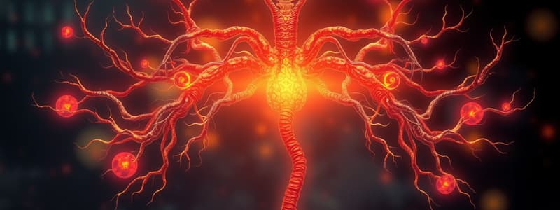
Communication Systems in the Human Body
- The human body operates two primary communication systems: the Nervous System and the Endocrine System.
Nervous System
- Communicates via electrochemical impulses and neurotransmitters.
- Regulates critical functions such as muscle contractions and glandular secretions.
- Provides a rapid response with effects lasting seconds and confined to localized areas.
Endocrine System
- Functions through chemical messengers called hormones, which are released into the bloodstream.
- Hormones influence a variety of processes including growth, development, reproduction, and metabolic activities.
- Actions are slower, taking minutes to weeks, and have widespread effects throughout the body.
Comparison of Systems
- The Nervous System is characterized by its quick action and short-term effects, while the Endocrine System operates slowly but facilitates long-lasting changes.
- Together, these systems work intricately to regulate bodily functions and maintain homeostasis.
Diagram Representation
- The Nervous System is illustrated by a red blood vessel with a purple signal, representing swift electrical impulses.
- The Endocrine System features yellow tissue showcasing a protein released by a gland along with a hormone directed towards target cells.
General Function of Endocrine Secretions
- Endocrine glands produce and secrete hormones, acting as chemical messengers in the bloodstream.
- Hormones travel through the blood to reach specific target cells, initiating various physiological responses.
Types of Hormone Receptors
- Cell surface receptors are associated with peptide hormones and catecholamines, influencing cell surface activities.
- Intracellular receptors interact with steroid and thyroid hormones, affecting gene expression and cellular function.
Role of Hormones
- Endocrine hormones play a crucial role in regulating:
- Growth and development processes in the body.
- Reproductive functions, including sexual development and reproductive health.
- Metabolic activities, encompassing both physical and chemical processes essential for homeostasis and energy production.
Pituitary Gland Overview
- The pituitary gland, commonly known as the hypophysis, is a crucial endocrine gland situated at the base of the brain.
- It regulates various bodily functions by releasing hormones that control other endocrine glands.
Anterior Pituitary
- Comprises about 75% of the pituitary gland's total mass.
- Produces hormones such as growth hormone (GH), prolactin (PRL), adrenocorticotropic hormone (ACTH), thyroid-stimulating hormone (TSH), and gonadotropins (LH and FSH).
- Hormone secretion is regulated by hypothalamic releasing and inhibiting hormones.
Posterior Pituitary
- The smaller posterior portion of the pituitary gland, primarily involved in storage and release.
- Stores two key hormones: oxytocin (involved in childbirth and lactation) and vasopressin (ADH, which regulates water balance).
- Does not produce hormones; it releases hormones synthesized in the hypothalamus.
Thalamus
- A critical relay station in the brain for sensory and motor signals.
- Also plays a role in regulating consciousness, sleep, and alertness.
- Not part of the pituitary gland but closely associated within the brain's functional network.
Hypothalamus
- A small region of the brain below the thalamus, crucial for homeostasis.
- Regulates body temperature, hunger, thirst, fatigue, and circadian rhythms.
- Produces releasing and inhibiting hormones that control the anterior pituitary's secretion.
Infundibulum
- A stalk-like structure connecting the hypothalamus to the pituitary gland.
- Serves as a conduit for neurohormones from the hypothalamus to reach the posterior pituitary.
- Plays an essential role in the communication between the hypothalamus and the pituitary gland.
Overview of the Hypophysis (Pituitary Gland)
- The pituitary gland, also known as the hypophysis, is a critical endocrine organ in the brain responsible for hormone regulation.
- It is located beneath the hypothalamus and is divided into anterior and posterior lobes.
Structural Components
- Optic Chiasma: The point where optic nerves cross, located near the pituitary gland.
- Pars Tuberalis: A tubular sheath extending from the pars distalis that encases the pituitary stalk, playing a role in hormone secretion.
- Pars Distalis: The anterior lobe of the pituitary gland, responsible for producing and releasing various hormones, including growth hormone and adrenocorticotropic hormone.
- Pars Intermedia: The intermediate lobe of the pituitary, which is particularly active in some animals but less so in humans, secreting melanocyte-stimulating hormone (MSH).
- Median Eminence: A part of the hypothalamus that forms a connection with the pituitary gland through the hypophyseal portal system, facilitating hormone transport.
- Infundibular Stem: A stalk that connects the pituitary gland to the hypothalamus, allowing for communication between the two.
- Pars Nervosa: The posterior lobe of the pituitary gland (neurohypophysis), which stores and releases hormones produced by the hypothalamus.
Anterior Hypophysis (Adenohypophysis)
- Composed of glandular tissue and regulates several hormones that influence growth, metabolism, and reproduction.
- Common hormones released include follicle-stimulating hormone (FSH), luteinizing hormone (LH), and thyroid-stimulating hormone (TSH).
Posterior Hypophysis (Neurohypophysis)
- Comprised mainly of nerve endings that originate from the hypothalamus.
- Stores and secretes hormones such as oxytocin and vasopressin (antidiuretic hormone), which are synthesized in the hypothalamus.
Ventral View of the Brain
- Frontal Lobe: Responsible for higher cognitive functions, decision-making, and voluntary movement.
- Temporal Lobe: Involved in processing auditory information and plays a crucial role in memory and emotion.
- Cerebellum: Coordinates voluntary movements and maintains posture, balance, and motor learning.
- Tuber Cinereum: Part of the hypothalamus, involved in endocrine functions and regulation of body temperature.
- Pons: Connects different parts of the brain, relaying messages between the cerebellum and the cortex.
- Anterior Median Fissure: A deep groove along the front of the spinal cord, aiding in the division of the right and left sides.
- Medulla Oblongata: Controls autonomic functions such as breathing, heart rate, and blood pressure.
- Hypophysis/Pituitary Gland: Often termed the "master gland," it regulates numerous physiological processes through hormone release.
- Mammillary Bodies: Linked to the limbic system, significant for memory processing and emotional responses.
- Pyramid: Houses motor fibers originating from the cortex that travel to the spinal cord.
- Decussation of Pyramids: Location where motor fibers cross over to contralateral side prior to reaching the spinal cord.
- 1st and 2nd Cervical Nerve: The first nerves emanating from the cervical region, crucial for head and neck movement.
- Optic Chiasm: The crossing point of optic nerves, enabling visual information processing from both eyes.
Sagittal View of the Brain
- Infundibulum: A stalk-like structure connecting the pituitary gland to the hypothalamus, involved in hormone transport.
- Median Eminence (Eminentia Mediana): Part of the hypothalamus that regulates hormone release into the pituitary gland.
- Mammillary Body: Plays a role in memory formation, particularly in the retrieval of memories.
- Basal Pons: Contains ascending and descending pathways, playing an essential role in the regulation of sleep, respiration, and swallowing.
- Pontine Tegmentum: A section that contains nuclei involved in motor control and sensory analysis, also crucial for regulating arousal and sleep.
Hypophysis/Pituitary Gland Overview
- The pituitary gland, also known as the hypophysis, is crucial for hormone regulation in the body.
- It consists of two main divisions: the anterior and posterior hypophysis.
Anterior Hypophysis
- Comprises three regions: pars tuberalis, pars distalis, and pars intermedia.
- Pars tuberalis: Forms a sheath around the infundibulum and is involved in the regulation of several hormones.
- Pars distalis: The largest portion, responsible for producing a variety of hormones including growth hormone, prolactin, and thyroid-stimulating hormone.
- Pars intermedia: Located between the other two segments, it produces melanocyte-stimulating hormone (MSH) which influences pigmentation.
Posterior Hypophysis
- Composed of three main parts: median eminence, infundibular stem, and pars nervosa.
- Median eminence: Acts as a critical communication point between the hypothalamus and pituitary, allowing for hormone release.
- Infundibular stem: The stalk connecting the pituitary gland to the hypothalamus, facilitating hormone transport.
- Pars nervosa: Stores and releases oxytocin and vasopressin (antidiuretic hormone) produced in the hypothalamus.
Development of Adenohypophysis and Neurohypophysis
- Development involves differentiation from two distinct ectodermal tissues: oral ectoderm (forming the adenohypophysis) and neural ectoderm from the diencephalon (leading to the neurohypophysis).
- Stippling indicates the oral ectoderm, while colored areas represent the neural ectoderm, visualizing their developmental origins.
Posterior Hypophysis
- Also known as the neurohypophysis, it is derived from neural tissue and is involved in the storage and release of hormones.
- Comprises the pars nervosa, which is the main tissue area where axonal endings release hormones into the bloodstream.
Anterior Hypophysis
- Referred to as the adenohypophysis, it functions as an endocrine gland, synthesizing and releasing various hormones.
- Contains the pars distalis, which is the largest portion of the adenohypophysis and responsible for producing key hormones involved in growth, metabolism, and reproduction.
Magnification Observations
- Low magnification: Provides an overview of the architecture and organization of both the posterior and anterior hypophysis.
- High magnification: Reveals cellular structures and types within the pars nervosa of the neurohypophysis and the pars distalis of the adenohypophysis, allowing for detailed observation of hormone-producing cells and their arrangement.
Overview of the Pituitary Gland
- Composed of two main parts: anterior hypophysis (adenohypophysis) and posterior hypophysis (neurohypophysis).
- Plays a crucial role in hormone production and regulation within the endocrine system.
Posterior Hypophysis (Neurohypophysis)
- Contains typical neurons that are integral to hormone secretion.
- Includes the pars nervosa, which is the larger part of the posterior pituitary.
- Responsible for the release of specific hormones, such as oxytocin and vasopressin (ADH), directly into the bloodstream.
Anterior Hypophysis (Adenohypophysis)
- Functions as a typical endocrine gland, involved in hormone synthesis and secretion.
- Comprises the pars distalis, which is the primary component of the anterior pituitary.
- Produces various hormones like growth hormone (GH), prolactin (PRL), and adrenal corticotropic hormone (ACTH) that regulate different physiological processes.
Microscopic Structure
- The pars nervosa consists of neuronal cells, while the pars distalis contains clusters of endocrine cells organized into glands.
- Histological differences between the two parts are significant for their functional roles in hormone production and secretion.
Posterior Hypophysis: Neurohypophysis
- Composed of typical neurons and the pars nervosa region.
- Pituicytes serve as glial cells, aiding in the storage and release of neurohypophysial hormones.
- Contains unmyelinated axons responsible for transmitting nerve impulses.
- Blood supply facilitated by endothelial cells of capillaries, ensuring efficient hormone delivery.
Anterior Hypophysis: Adenohypophysis
- Functions as a typical endocrine gland with various hormone-secreting cells.
- Pars distalis is the main secretory portion.
- Acidophils (pink): secrete polypeptide hormones including:
- Growth Hormone (GH): crucial for growth and metabolism.
- Prolactin (PRL): key in lactation and reproductive functions.
- Basophils (dark purple): secrete glycoprotein hormones such as:
- Corticotrophin (ACTH): stimulates adrenal cortex to release cortisol.
- Thyroid Stimulating Hormone (TSH): regulates thyroid gland activity and metabolism.
- Gonadotrophins (FSH & LH): control reproductive processes and hormone production in ovaries and testes.
- Chromophobes (pale): appear depleted of hormones with scant cytoplasmic granules; may exhibit some secretory activity.
Blood Supply to the Anterior Lobe of Adenohypophysis
- Blood is supplied to the adenohypophysis via the superior hypophyseal arteries, which are branches of the internal carotid artery.
- A primary plexus forms at the base of the hypothalamus, allowing the initial vascular network.
- Blood drains from this primary plexus through hypophyseal portal vessels to a secondary plexus located in the anterior lobe.
- Venous drainage occurs through anterior inferior hypophyseal veins, completing the circulatory route.
Key Components of Blood Supply
- Capillaries in the median eminence play a critical role in the vascular network.
- The superior hypophyseal arteries are crucial for initial blood supply.
- Long portal vessels facilitate the connection between the primary and secondary vascular plexuses.
- Capillaries in the pars distalis are integral for nutrient exchange.
- The inferior hypophyseal vein is involved in the outflow of blood from the anterior lobe.
Structural Parts of the Anterior Lobe
- The anterior lobe consists of three distinct regions:
- Pars nervosa
- Pars distalis
- Pars intermedia
- Each part has unique functions and contributions to overall hormone regulation and secretion in the body.
Blood Supply: Posterior Lobe - Neurohypophysis
- Supplied by inferior hypophyseal arteries, which branch from the internal carotid artery.
- Contains a vascular plexus located within the posterior lobe, essential for hormone release.
- Venous drainage of the posterior lobe occurs through posterior inferior hypophyseal veins leading to the cavernous sinus.
Structure
- Pars Nervosa:
- Houses a capillary plexus crucial for hormone exchange.
- Pars Distalis:
- One of the primary sections of the pituitary gland, important for hormone production.
- Inferior Hypophyseal Veins:
- Drains blood from the inferior hypophyseal arteries after nutrient exchange.
- Inferior Hypophyseal Arteries:
- Supply blood directly to the neurohypophysis, facilitating delivery of essential hormones.
Hormones of the Pars Distalis
-
Growth Hormone (GH):
- Secreted by somatotropes.
- Targets liver and adipose tissue.
- Stimulates growth and metabolism of carbohydrates and lipids.
-
Prolactin (PRL):
- Released by lactotropes.
- Targets mammary glands.
- Essential for milk production.
-
Thyroid Stimulating Hormone (TSH):
- Produced by thyrotropes.
- Targets the thyroid gland.
- Promotes secretion of thyroid hormones, influencing metabolism.
-
Follicle Stimulating Hormone (FSH):
- Secreted by gonadotropes.
- Targets ovaries and testes.
- Regulates reproductive functioning, including oocyte maturation and spermatogenesis.
-
Luteinizing Hormone (LH):
- Also produced by gonadotropes.
- Targets ovaries and testes.
- Stimulates production of sex hormones, such as estrogen and testosterone.
-
Adrenocorticotropic Hormone (ACTH):
- Released by corticotropes.
- Targets adrenal gland (cortex).
- Stimulates secretion of glucocorticoids, involved in stress response and metabolism.
-
β-Endorphin:
- Produced by corticotropes.
- Targets opioid receptors.
- Acts to inhibit pain perception, contributing to the body's pain relief system.
Pars Nervosa Cells & Hormones
- Pars nervosa is part of the posterior pituitary gland and releases neurohormones received from the hypothalamus.
- Antidiuretic Hormone (ADH), also known as vasopressin, is crucial for regulating water levels in the body, promoting water reabsorption in the kidneys.
- Oxytocin plays a significant role in stimulating uterine contractions during childbirth and facilitates milk ejection during breastfeeding.
- Both hormones are synthesized in the hypothalamus and transported to the pars nervosa for storage and release into the bloodstream.
Hypophysis: Clinical Imbalance of GH
- Hypopituitarism: Characterized by insufficient hormone production from the pituitary gland, leading to various clinical symptoms.
- Progeria: A rare genetic disorder resulting in accelerated aging; directly linked to hypopituitarism.
Hyperpituitarism
- Acromegaly: Condition caused by excessive growth hormone (GH) production, leading to abnormal growth in bone and soft tissue, particularly in hands, feet, and jaws.
- Giantism: Occurs during childhood before growth plates close; results in excessive height and size due to prolonged exposure to increased GH levels.
- Dwarfism: Can also be associated with an imbalance in GH, often linked to insufficient production during childhood, affecting overall growth and stature.
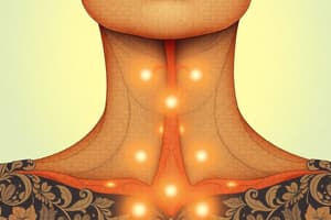
Structure of the Thyroid Gland
- Encased in a fibrous capsule for protection.
- Comprised of two distinct lobes: left and right.
- Lobes are joined by a tissue band called the isthmus.
Isthmus Location
- Positioned between the 2nd and 4th tracheal rings, closer to the lower section of the thyroid gland.
Anatomy Components
- Larynx: The voice box located above the thyroid.
- Thyroid Gland: Endocrine gland responsible for hormone production.
- Isthmus: The narrow connection between the thyroid lobes.
- Anterior View: Observation perspective showing the thyroid's front structure.
Microscopic Anatomy
- Colloid: A gel-like substance within the follicles that stores thyroid hormones.
- Follicular Cells: Cells that line the thyroid follicles and synthesize thyroxine (T4) and triiodothyronine (T3).
- Extrafollicular Cells: Cells involved in producing calcitonin, which helps regulate calcium levels in the blood.
Structure of the Thyroid Gland
- Comprised of multiple structures working together to regulate metabolism and calcium homeostasis.
Follicles
- The basic functional units of the thyroid gland, each surrounded by connective tissue.
- Contains colloid, a viscous fluid storing thyroid hormones.
- Reticular fibres provide structural support between the follicles.
- Each follicle is supplied with a capillary network for hormone transport and nutrient supply.
- Enclosed by a basal lamina, which acts as a selective barrier.
- Follicular cells line the follicles and are responsible for the secretion of thyroid hormones (TH), primarily thyroxine (T4) and triiodothyronine (T3).
Parafollicular Cells (C Cells)
- Located between the thyroid follicles.
- Secrete calcitonin, a hormone involved in regulating calcium levels in the blood by lowering them when necessary.
Inactive Thyroid Cells
- Characterized by low, simple (flat) cuboidal epithelial cells
- Follicles contain colloid, a substance that stores thyroid hormones
- Colloid is rich in thyroglobulin, which serves as the storage form for T3 (triiodothyronine) and T4 (thyroxine) hormones
Active Thyroid Cells
- Comprised of tall, simple cuboidal epithelial cells
- Follicles still contain colloid but show reabsorption lacunae, appearing as white spaces or scallops
- Follicular cells actively extract stored thyroid colloid, converting it into bioactive T3 and T4 hormones
- Active hormones are then released into nearby capillaries, facilitating their distribution in the bloodstream
Thyroid Gland and Hormones
- The thyroid gland synthesizes and releases thyroid hormones (TH).
- Main hormones produced:
- Thyroxine (T4)
- Triiodothyronine (T3)
Functions and Targets
- Regulates basal metabolic rate affecting:
- Heart rate
- Blood pressure
- Body temperature
- Essential for normal development in:
- Nervous system
- Skeleton
- Reproductive systems
- Influences metabolism of carbohydrates, lipids, and proteins.
- Affects nearly all body systems apart from:
- Brain
- Some gonads
- Spleen
- The thyroid gland itself
Stimulation of Thyroid Gland
- Thyroid activity is stimulated by:
- Blood levels of thyroxine (T4)
- Thyroid Stimulating Hormone (TSH)
Regulation Mechanism
- Hypothalamus releases Thyrotropin-releasing hormone (TRH) into the bloodstream.
- TRH prompts the pituitary gland to secrete TSH.
- TSH stimulates the thyroid gland to produce T3 and T4.
- Levels of T3 and T4 in bloodstream provide feedback to the hypothalamus and pituitary, regulating TRH and TSH production.
T3 vs T4
- T3 acts more quickly and has a shorter duration of action but is produced in smaller amounts compared to T4.
- This feedback loop maintains appropriate levels of thyroid hormones to support normal bodily functions.
Thyroid Gland Functions
- Produces and secretes calcitonin, a hormone crucial for calcium regulation.
- Targets blood calcium and phosphate levels, ensuring equilibrium within the body.
- Stimulates growth in long bones, contributing to overall skeletal health.
- Lowers blood calcium (Ca++) levels, contrasting the effects of parathyroid hormone (PTH).
Hormonal Antagonism
- Calcitonin serves as an antagonist to parathyroid hormone (PTH).
- PTH regulates calcium and phosphate metabolism by increasing blood Ca++ levels, thus calcitonin counters this effect.
- Inhibits osteoclast activity, the cells responsible for bone resorption, reducing the release of calcium into the bloodstream.
Regulatory Mechanism
- Calcitonin secretion is controlled by the levels of calcium in the blood vessels.
- Higher calcium levels trigger the release of calcitonin to promote bone deposition and reduce circulating calcium.
Mechanism of Action
- Calcitonin inhibits osteoclasts, reducing their activity and therefore lowering blood calcium levels.
- It also facilitates the deposition of calcium salts in the bones, enhancing bone strength and density.
Hyperthyroidisms
- Characterized by overactivity of the thyroid gland leading to excessive production of thyroid hormones.
- Often referred to as thyrotoxicosis; a common cause is Graves' disease, an autoimmune disorder.
- Symptoms can range from mild (asymptomatic) to severe, including a life-threatening condition known as thyroid storm.
Hypothyroidisms
- Defined by underactivity of the thyroid gland, resulting in insufficient thyroid hormone production.
- Associated conditions include:
- Goitre: enlargement of the thyroid gland, often due to iodine deficiency.
- Cretinism: severe hypothyroidism in infancy or childhood, leading to stunted physical and mental development.
- Myxedema: a serious form of hypothyroidism in adults, characterized by extreme swelling and metabolic slowdown.
- Hashimoto's thyroiditis: an autoimmune disease causing chronic inflammation of the thyroid, leading to gradual loss of function.
General Notes
- Both hyperthyroidisms and hypothyroidisms can result in notable clinical signs and symptoms, impacting metabolic rate and overall health.
- Monitoring and treatment of thyroid hormone levels are critical for managing both clinical states effectively.
Anatomy of the Parathyroid Gland
- Size of the parathyroid gland is approximately 6x3x2 mm.
- Color of the gland is yellow-brown.
- Composed of two pairs of glands: two superior and two inferior.
Functions and Location
- Positioned on the posterior aspect of the thyroid gland.
- Regulates calcium levels in the bloodstream, critical for various physiological processes.
- Secretes parathyroid hormone (PTH), which elevates calcium levels in the blood.
Conditions Related to Parathyroid Function
- Hypoparathyroidism occurs when there is insufficient production of PTH, leading to low calcium levels.
- Hyperparathyroidism is characterized by excessive PTH production, resulting in high calcium levels.
Additional Facts
- Approximately 5% of individuals may have more than four parathyroid glands, indicating anatomical variability.
Parathyroid Gland
- Small, oval-shaped gland situated on the posterior side of the thyroid gland.
- Surrounded by a thin layer of connective tissue known as the capsule, which protects and supports the gland.
- Contains fenestrated capillaries that feature pores, facilitating the exchange of substances between blood and glandular tissue.
- Composed of reticular fibers that provide structural support to the gland's cells.
- Glandular cells are the primary cell type within the parathyroid, responsible for the synthesis of parathyroid hormone (PTH), crucial for calcium homeostasis.
- Divided into lobules by septa, which are small partitions of connective tissue, enhancing compartmentalization and functional organization.
Thyroid
- Located in the neck, beneath the Adam's apple, and plays a key role in metabolism regulation.
- Produces several hormones, including thyroxine (T4) and triiodothyronine (T3), which significantly impact energy regulation, growth, and development.
Histology
- Involves the study of tissue structure through thinly sliced and stained tissue samples, allowing microscopic examination of cellular composition and arrangement.
Parathyroid Gland Structure and Composition
- Chief Cells: Dark purple cells responsible for producing parathyroid hormone (PTH), crucial for regulating calcium levels in the body.
- Oxyphil Cells: Larger, pale cells with an unidentified function, appearing in the gland around puberty.
- Adipose Tissue: Fat cells that start to appear at puberty, gradually increasing in quantity over time, suggesting a change in gland composition.
- Blood Vessel: Integral for providing oxygen and nutrients, as well as transporting hormones produced by the parathyroid gland.
- Connective Tissue Septum: Structure that divides the gland into lobules, supporting the overall integrity and organization of the gland.
Cells in the Parathyroid Gland
- Oxyphil cells are large, pale cells characterized by a significant amount of cytoplasm, which distinguishes them from other cell types.
- Chief cells are smaller, darker, and appear purple under microscopic examination; they play a crucial role in hormone production.
- Adipose tissue in the parathyroid gland consists of fat cells, identifiable by their clear cytoplasm and large size.
- The parathyroid gland contains a mix of oxyphil cells, chief cells, and adipose tissue, which together contribute to its function and structure.
- The classification of chief cells as darker and the visibility of different cell types depend on the quality of the microscopic image and its interpretation.
Parathyroid Gland Overview
- Produces parathyroid hormone (PTH), critical for calcium regulation.
- PTH acts as an antagonist to calcitonin, which is produced by the thyroid gland.
Functions of Parathyroid Hormone
- Regulates blood calcium and phosphate levels.
- Increases blood calcium levels by stimulating osteoclast activity.
- Inhibits calcium uptake in the kidneys.
- Plays a role in calcium (Ca²⁺) and phosphate (P) metabolism.
Regulation Mechanism
- Blood vessel calcium levels directly control PTH secretion.
- Elevated calcium levels in the blood lead to lower PTH production.
Calcium Homeostasis
- Blood calcium homeostasis maintained between 9-11 mg/100 ml.
- PTH promotes osteoclast activity, leading to increased blood calcium concentration.
- Calcitonin encourages calcium salt deposits in bones, lowering blood calcium levels.
Calcium Homeostasis
- Blood calcium levels are maintained within a narrow range of 9-11 mg/100 ml to ensure physiological balance.
- Increased blood calcium levels are facilitated by:
- Enhanced absorption of calcium in the kidneys.
- Release of calcium from bones into the bloodstream.
- Increased production of calcitriol, which boosts calcium absorption in the digestive system.
- The thyroid gland secretes calcitonin, which:
- Inhibits osteoclast activity, decreasing bone resorption.
- Leads to a reduction in blood calcium levels.
- Elevated calcium levels promote calcium deposition in bones, promoting bone health.
- Decreased blood calcium levels trigger the release of parathyroid hormone (PTH) from the parathyroid glands.
- PTH stimulates osteoclasts, resulting in increased calcium release from bones, which raises blood calcium levels.
- A feedback mechanism exists where calcitonin and PTH work antagonistically to regulate calcium levels, enhancing homeostasis in the body.
Hyperparathyroidism Symptoms
Digestive System
- Symptoms include loss of appetite, nausea, vomiting, and constipation.
Nervous System
- Patients may experience fatigue, depression, and confusion.
Musculoskeletal System
- Symptoms manifest as muscle weakness and aches or pains in bones and joints.
Urinary System
- Common issues are kidney stones, increased thirst, and increased urination.
Hormonal Imbalance Effects
- Elevated phosphate (P) levels with decreased calcium (Ca2+) can cause nervous excitability, involuntary muscle contractions, and may lead to death.
- High calcium (Ca2+) levels with low phosphate (P) can result in weakened bones and the formation of kidney stones.
Studying That Suits You
Use AI to generate personalized quizzes and flashcards to suit your learning preferences.
Description
Test your knowledge on the various types of endocrine glands and organs that contribute to the endocrine system. This quiz covers key structures such as the pineal gland, pituitary gland, and the role of other organs like the pancreas and gonads. Challenge yourself on the functions and characteristics of these crucial components in hormone production.