Podcast
Questions and Answers
What is the primary purpose of clearing in tissue processing?
What is the primary purpose of clearing in tissue processing?
- To enhance cell viability
- To give tissue a translucent appearance
- To increase the tissue stiffness
- To replace ethanol with paraffin (correct)
Which components are stained by basic dyes?
Which components are stained by basic dyes?
- Nucleic acids and glycosaminoglycans (correct)
- Lipids and carbohydrates
- Actin filaments and collagen
- Cell membranes and nuclei
Which statement regarding paraffin embedding is accurate?
Which statement regarding paraffin embedding is accurate?
- It is suitable only for electron microscopy.
- Paraffin embeds using cross-linking polymerizers.
- It requires temperatures over 100°C.
- Infiltration with paraffin avoids tissue distortion. (correct)
What does hematoxylin primarily stain in tissue sections?
What does hematoxylin primarily stain in tissue sections?
What is the typical thickness of paraffin sections cut for light microscopy?
What is the typical thickness of paraffin sections cut for light microscopy?
Flashcards are hidden until you start studying
Study Notes
Tissue Preparation
- Ethanol is used to dehydrate tissue samples, replacing water with an organic solvent.
- This process is called clearing as the tissue becomes translucent.
- The dehydrated tissue is then infiltrated with paraffin, which is melted at 52°C-60°C, creating a hard embedding medium.
- Plastic embedding avoids the high temperatures of paraffin, helping to prevent tissue distortion.
- The embedded tissue is sectioned on a microtome, typically producing sections 3-10 μm for light microscopy, and thinner sections for electron microscopy.
Staining
- Staining allows for the visualization of specific cell structures.
- Basophilic components of cells, such as DNA and RNA, have a high affinity for basic dyes.
- Examples of basic dyes include toluidine blue, alcian blue, and methylene blue.
- Acidophilic components, such as proteins with many ionized amino groups, bind readily with acidic dyes.
- Examples of acidic dyes include eosin, orange G, and acid fuchsin.
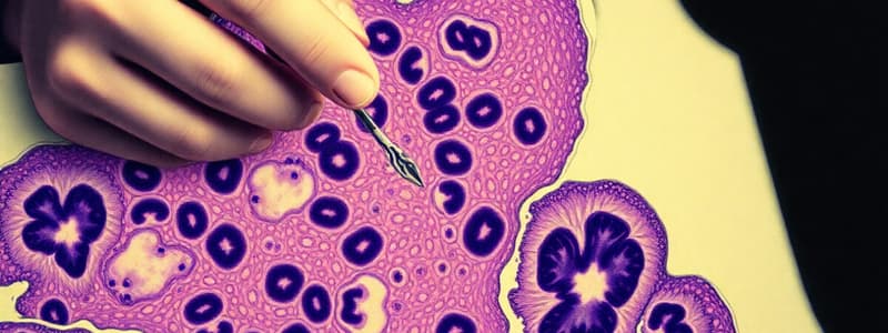
- The most common staining method is Hematoxylin and Eosin (H&E).
- Hematoxylin stains DNA in the cell nucleus, RNA-rich portions of the cytoplasm, and the matrix of cartilage, producing a dark blue or purple color.
- Eosin stains cytoplasmic structures and collagen pink.
Microscopy
- Bright-field microscopy requires staining to visualize cells.
- Phase-contrast microscopy enhances contrast of unstained cells, allowing for observation of live cells.
- Differential interference contrast microscopy (DIC) creates a 3D-like image of cells and is also used for live cells.
- Polarized light microscopy can visualize birefringent structures, which have the ability to rotate the direction of polarized light.
Other Techniques
- Autoradiography uses radiolabeled substances to track the location and movement of molecules in cells and tissues.
- Enzyme histochemistry localizes cellular structures using specific enzyme activities, often using unfixed or mildly fixed tissue sectioned on a cryostat.
- Immunocytochemistry uses antibodies labeled with fluorescent dyes, enzymes, or other markers to detect specific proteins or antigens in cells and tissues.
- In situ hybridization techniques detect specific nucleotide sequences in DNA or RNA in cells and tissues.
- Cell and tissue culture allows for the study of living cells and tissues outside of the body, providing a controlled environment for experiments.
Studying That Suits You
Use AI to generate personalized quizzes and flashcards to suit your learning preferences.


