Podcast
Questions and Answers
What is the total preparation time for light microscopy?
What is the total preparation time for light microscopy?
- 12 hours to 2½ days (correct)
- 3 days to 1 week
- 30 minutes to 24 hours
- 1 hour to 5 hours
Which type of microscopy does not require staining to view the slide?
Which type of microscopy does not require staining to view the slide?
- Phase-contrast microscopy (correct)
- Fluorescence microscopy
- Bright-field microscopy
- Polarizing light microscopy
What is the immediate frozen section time for results in tumor surgery?
What is the immediate frozen section time for results in tumor surgery?
- 15 minutes
- 30 minutes
- 5 minutes (correct)
- 1 minute
What step immediately follows fixation when preparing a tissue sample?
What step immediately follows fixation when preparing a tissue sample?
Which type of microscopy is most commonly used by students and pathologists?
Which type of microscopy is most commonly used by students and pathologists?
What color does hematoxylin impart to the nuclei of cells?
What color does hematoxylin impart to the nuclei of cells?
What does eosin primarily stain in tissue sections?
What does eosin primarily stain in tissue sections?
Which stain preserves mucous in goblet cells and stains it magenta?
Which stain preserves mucous in goblet cells and stains it magenta?
What type of components does Toluidine blue stain specifically in tissues?
What type of components does Toluidine blue stain specifically in tissues?
Which stain would you use to visualize lipids in frozen sections?
Which stain would you use to visualize lipids in frozen sections?
What type of fibers does silver impregnation stain?
What type of fibers does silver impregnation stain?
Which stain is primarily used for blood smears and diagnosing diseases like malaria?
Which stain is primarily used for blood smears and diagnosing diseases like malaria?
Metachromasia is most prominently observed in which of the following?
Metachromasia is most prominently observed in which of the following?
What is the primary focus of histology?
What is the primary focus of histology?
Which of the following is NOT a step in the preparation of histological sections?
Which of the following is NOT a step in the preparation of histological sections?
What is the function of the fixation step in tissue preparation?
What is the function of the fixation step in tissue preparation?
What is the common thickness for sections cut for light microscopy?
What is the common thickness for sections cut for light microscopy?
Which type of tissue is characterized by having a high degree of cellular structure?
Which type of tissue is characterized by having a high degree of cellular structure?
Which staining process is used to identify specific structures and molecules in histological sections?
Which staining process is used to identify specific structures and molecules in histological sections?
What is the role of the extracellular matrix in tissues?
What is the role of the extracellular matrix in tissues?
What is commonly used as a fixative to preserve tissue samples?
What is commonly used as a fixative to preserve tissue samples?
Flashcards are hidden until you start studying
Study Notes
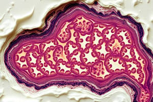
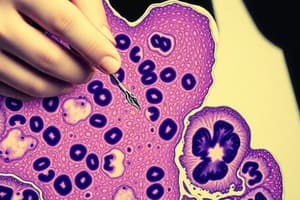
Histology Overview
- Definition: Histology is the study of the microscopic anatomy of tissues and cells.
- Origin of the term: Derived from Greek "Histos" meaning tissue, and "logos" meaning study.
- Techniques: Involves examining thin slices of tissue with light or electron microscopes using various histological stains.
Levels of Biological Organization
- Cells: The smallest independently functioning units, highly complex structures.
- Tissues: Groups of similar cells sharing functions and origin.
- Organs: Composed of multiple tissue types (e.g., liver, lungs, kidneys).
- Systems: Combinations of organs working together for specific functions (e.g., digestive, respiratory, urinary systems).
Tissue Composition
- Made up of cells and extracellular matrix.
- Four primary types: Epithelial, connective, muscular, and nervous tissues.
Steps in Tissue Preparation
- Fixation: Preserves tissue shape and volume, prevents autolysis; formaldehyde is a common fixative.
- Processing:
- Dehydration: Removes fixative and water, replacing with ethanol.
- Clearing: Uses xylene to remove alcohol.
- Embedding: Tissue embedded in wax for support during sectioning.
- Cutting: Microtome slices tissue into sections (2 to 25 micrometers for light microscopy, 60 to 100 nanometers for electron microscopy).
- Staining: Uses various dyes to highlight specific structures.
- Mounting: Covers sections with a thin glass cover-slip.
Staining Techniques
- Hematoxylin and Eosin (H&E): Universal routine stain; hematoxylin (blue) stains acidic cell components; eosin (red) stains basic components.
- Periodic acid-Schiff (PAS) Staining: Highlights mucus and glycogen; stains goblet cells magenta.
- Toluidine Blue: Stains specific tissue components with metachromasia, appearing purple.
- Oil Red O: Stains lipids red-orange in frozen sections.
- Sudan Black: Stains lipids black in frozen sections.
- Silver Impregnation: Stains reticular fibers black.
- Giemsa Stain: Used for blood smears, helpful in diagnosing diseases like malaria.
Types of Microscopy
- Light Microscopy:
- Bright-field: Most common, used for general observations.
- Phase-contrast: Does not require staining; ideal for observing living cells.
- Polarizing: Highlights tissue structures against a dark background.
- Fluorescence: Shows only fluorescent molecules under UV light.
- Electron Microscopy:
- Transmission Electron Microscopy (TEM): Produces high-resolution images at ultrastructural levels.
- Scanning Electron Microscopy (SEM): Provides 3D surfaces of samples.
Preparation Time
- Total preparation for light microscopy: Takes between 12 hours to 2.5 days.
- Immediate frozen section preparation for quick results: Takes about 5 minutes, useful in tumor surgery.
Case Scenario
- A biopsy sample is fixed using formaldehyde; the next step is dehydration as part of the tissue processing sequence.
Studying That Suits You
Use AI to generate personalized quizzes and flashcards to suit your learning preferences.